
Web-Based Viewing Intro
With just a browser and an internet connection, you may use a web-based DICOM viewer for Windows, such Post DICOM, the OHIF Viewer, or 3DICOM Patient Viewer, to remotely examine 3D DICOM Model Images on any Windows device. On any contemporary Windows machine, you may access sophisticated 3D medical visualization tools by uploading your DICOM data.
Alternatively, a few structures OF 3D DICOM Model Images offer downloadable patron packages, inclusive of 3D Slicer, which offer more advanced equipment for in-depth evaluation and 3D visualization.
Using a Web-Based DICOM Viewer
- This method, which uses a DICOM online viewer in your browser and doesn’t require installation, is the most practical approach to examine 3D DICOM Model Images remotely.
1. Choose a Web-Based Platform:
- Select a platform like:
- Post DICOM: Offers superior functions, along with 3D DICOM Model Images rendering and photo annotation, for professional use.
- 3D Slicer: An open-source platform for visualizing and analyzing medical images, which can also be used for 3D visualization.
- IMAIOS DICOM Viewer: A free, web-based software to open and view 3D DICOM Model Images documents without delay for your browser.
- OHIF Viewer: A customizable, open-source viewer that integrates with present healthcare systems.
2. Upload Your DICOM Files:
- Once you’ve accessed the platform, upload your DICOM documents from your Windows device.
3. View in 3D:
- The platform’s interface will then system the photos and let you view them in 3D DICOM Model Images.
- Considerations
Internet Connection:
A solid internet connection is important for using internet-primarily based visitors.
1. File Storage:
Make sure you are familiar with the platform’s security and data privacy policies because some web systems could also let you upload your papers to the network. The ALM DICOM Viewer’s Zero Trust Security architecture guarantees compliance-ready protection of medical data in both on-site and cloud contexts for optimal security.
2. Advanced Features:
For complex analysis or primary diagnostic use, keep in mind platforms like MedDream, which is FDA cleared and gives diagnostic features inside a web browser environment.
Main Techniques:
There are two main techniques for remotely viewing 3-D dicom images on any Windows tool: using an internet-based “Zero Footprint” viewer or a computing device viewer related to a Picture Archiving and Communication System (PACS).
Method 1: Use an internet-based 3D DICOM Model Images viewer (Zero Footprint)
This is the most practical and efficient approach. Without the need to install additional software, a Zero Footprint DICOM viewer may be accessed from practically any Windows device by running directly in a web browser. The ALM DICOM Viewer Hybrid Model offers smooth browser-based access with enterprise-grade functionality, which is exactly what many healthcare professionals want.
How it works
- Access the server: The 3D DICOM Model Images photographs are hosted on a server (PACS or cloud). You are furnished a stable hyperlink to get access to the viewer through your web browser.
- View from everywhere: Once you have the URL and credentials, you can open the viewer on any Windows PC with an internet connection and an internet browser.
- Perform three-D rendering: The server handles the heavy processing, so you can perform three-D capabilities like Maximum Intensity Projection (MIP), Multiplanar Reconstruction (MPR), and extent rendering directly inside the browser, even on a low-electricity pc.
Popular 0-footprint viewers
- OHIF Viewer: An open-source, web-based DICOM viewer for Windows that works with a variety of image archives and is quite customisable.
- MedDream DICOM Viewer: A for-profit online viewer with sophisticated 3D medical imaging capabilities that interfaces with PACS systems.
- RemotEye Viewer: Neologica’s browser-based RemotEye Viewer is capable of producing sophisticated 3D DICOM images.
Method 2: Use a computer 3D DICOM Model Images viewer with PACS
This technique entails setting up a specific Windows DICOM viewer on your device and configuring it to establish a connection with a distant PACS server. Although it has to be set up on each Windows computer, this approach provides full control and sophisticated functionality via the ALM DICOM Viewer On-Site Model, making it perfect for medical facilities and research centers that require safe local deployment.
How it works
- Install consumer software: You download and set up a 3D DICOM Model Images viewer utility, for your Windows computer.
- Configure network get admission to: You have to configure the viewer with the perfect IP address, port, and Application Entity (AE) title to connect to the PACS server.
- Query and retrieve pics: Once related, the viewer can search for and retrieve specific patient research from the server to show them locally.
- Perform 3D rendering: Many contemporary desktop viewers, such as 3D DICOM Model Images, offer robust 3D reconstruction capabilities, including MPR and extended rendering for downloaded studies.
Popular computer visitors for Windows
- 3D DICOM Model Images Viewer: A well-liked, quick, and user-friendly desktop viewer for Windows that provides 3D MPR and volume rendering.
- MicroDicom: A free and easy-to-use DICOM viewer for Windows that offers all the necessary processing, exporting, and viewing features.
- Weasis: A free, stand-alone, web-based program for viewing medical images that works with PACS systems and supports Windows.
To view DICOM (Digital Imaging and Communications in Medicine):
The dicom image viewer demo is on a PC, you can use a specialised software program designed for coping with clinical imaging documents.
Here are numerous options and steps to do so:
Software Options
1. DICOM Viewers:
- – OsiriX: A famous viewer for macOS; however, there is a version known as OsiriX Lite for Windows.
- – MicroDicom: A lightweight DICOM viewer for Windows with a person-friendly interface.
- DICOM Viewer: A rapid and easy-to-use viewer for Windows that helps view a couple of DICOM files.
- – Ginkgo CADx: An open-source DICOM viewer available on a couple of platforms.
- – Weasis: A pass-platform DICOM viewer that may be run as a standalone software or inside a web browser.
2. Image Editing Software:
- ImageJ:
A public domain Java-based picture processing program that can open 3D DICOM Model Images files with the best plugins.
- dicom web viewer:
Without the need for extra software, you may upload and view 3D DICOM Model Images straight from your web browser with certain online DICOM web viewer platforms.
Steps to View DICOM Images
1. Download and Install a DICOM Viewer:
– Choose one of the software alternatives listed above and download it from a reputable website.
– Follow the setup commands to set it up for your PC.
2. Open DICOM Files:
– Launch the 3D DICOM Model Images viewer.
– Use the “Open” or “Import” choice within the software to load your DICOM documents. You can usually choose out a couple of documents or a folder containing DICOM snapshots.
3. Navigate and Analyze:
– Once the photographs are loaded, you can use the software program’s gear to navigate through the photos, regulate evaluation/brightness, zoom in/out, and take a look at the photos as desired.
Save or Export:
– If you want to shop or export the snapshots in any other format (like JPEG or PNG), most visitors provide an export characteristic.
Tips
- Ensure that your 3D DICOM Model Images files are not corrupted and are in the right layout.
- Familiarize yourself with the features of the viewer you select, as they can vary substantially in terms of functionality and person interface.
1. Supports a couple of DICOM record sorts
The software can open and show research obtained from specific imaging modalities:
- Digital Radiography (CR, DX)
- Mammography (MG)
- Computed Tomography (CT)
- Magnetic Resonance (MR)
- Positron Emission Tomography (PET)
- Ultrasonography (US)
- Digital Angiography (XA)
- Gamma Camera, Nuclear Medicine (NM)
- Secondary Pictures and Scanned Images (SC)
- Structured Reports (SR)
2. Many styles of DICOM photos are supported:
Monochromatic (e.G. CR, CT, MR) and coloration (e.G. US, three-D reconstructions)
Static photographs (e.G. CR, MG, CT) and dynamic sequences (e.G. XA, US)
3. Basic gear for the manipulation of pictures
-
All the essential gear is near at hand
- DICOM Viewer provides the following fundamental gear for the manipulation and dimensioning of snapshots:
a) Fluid zooming and panning
- Brightness and contrast modifications, negative mode
- Preset window settings for Computed Tomography (lung, bone, and so forth.)
b) Segment duration
- Mean, minimal and most parameter values (e.G. Density in Hounsfield Units in Computed Tomography) inside circle/ellipse and its location
- Angle value (regular and Cobb perspective)
4. Pen device for freehand drawing
-
Lightning fast overall performance
- DICOM Viewer is designed to use resources as efficiently as possible. It can employ a multiprocessor and multicore system with huge quantities of gigabytes of RAM, but will even run on an old single-core gadget with handiest 1GB RAM.
Quick as lightning
DICOM Viewer was designed to apply sources as efficiently as possible. It can make use of a multiprocessor and multicore machine with huge amounts of gigabytes of RAM, but may also run on an old single-middle device with the simplest 1GB RAM.
A sixty-four-bit version is provided for contemporary structures to maintain all opened photographs in more than 4GB of memory, if necessary. Asynchronous analyzing helps you to browse and technique feature photos while they’re still being opened.
All of that is available in a single, very compact software that has an installer size of just over 7MB.
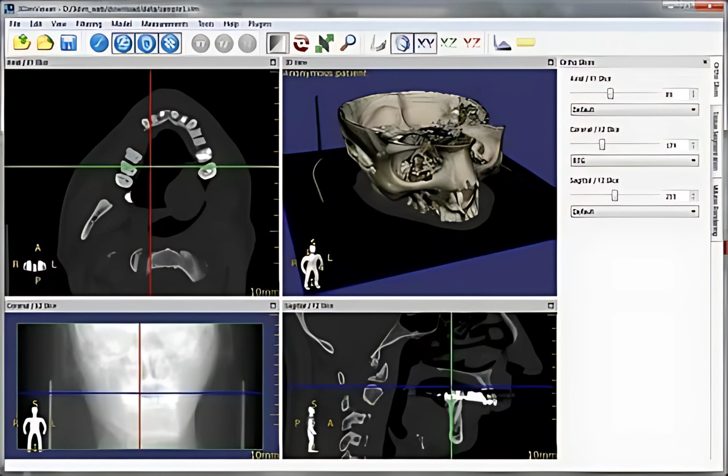
1. Browse and evaluate multiple series of pix
Compare different collections or research
Multiple collections of one look at or numerous studies can be simultaneously opened within the same or unique home windows for assessment purposes.
Series inclusive of snapshots which have been acquired within the same aircraft (e.G. Computed Tomography series earlier than and after management of the assessment medium) are mechanically synchronized by default.
Cross-reference strains are displayed for higher correlation of the anatomy while browsing the collection with exclusive picture planes (e.G. Magnetic Resonance Imaging).
2. PACS purchaser characteristics for looking at and retrieving studies
Search and download research from PACS locations
The PACS (Picture Archiving and Communication System) consumer feature we could use is 3D DICOM Model Images Viewer to question and retrieve research from other PACS hosts.
Supported provider elegance customers/vendors are: C-ECHO SCU, C-ECHO SCP, C-FIND SCU, C-MOVE-SCU, C-STORE-SCP.
Received 3D DICOM Model Images files may be saved in a temporary folder and deleted when it closes, or they may be saved inside the neighborhood database.
3. Local archive
The local archive feature lets you import DICOM studies from CD/DVDs, USB flash drives, local and network folders, or PAC
The neighborhood archive can be beneficial in situations in which the original media is not to be had, otherwise you would love to open the observe without the need to repeatedly retrieve it from a PACS server.
You can also access the database to organize and quickly locate studies for your collection of DICOM documents that are saved at the tough force.
4. Export DICOM files to BMP, JPEG, WMV
Export DICOM files to snapshots and movies
Create visually lovely displays and expert publications – DICOM Viewer can export DICOM documents to JPEG (compressed) or BMP (uncompressed bitmap) photographs and WMV (Windows Media Video) movies.
One photo, an entire collection or all open snapshots can be exported concurrently.
Displayed snapshots may be speedy copied to the Windows clipboard the usage of the CTRL C shortcut and may be fast and effortlessly pasted into Word or PowerPoint files.
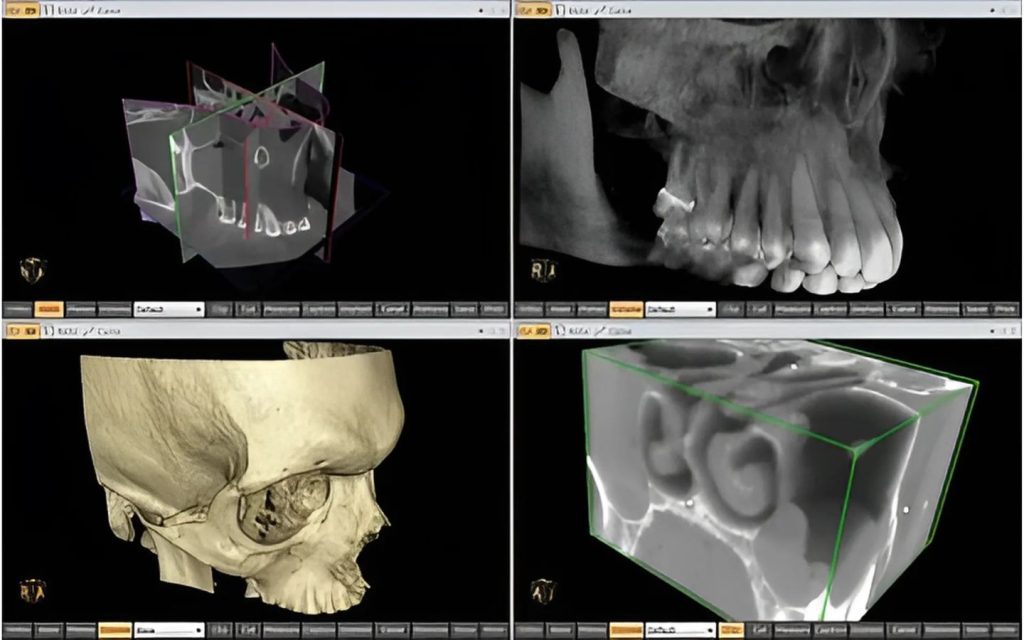
5. 2D and 3D Multi-planar reconstructions (MPR)
Multiplanar reconstructions
The MPR device supplied within DICOM Viewer may be used to reconstruct pics in orthogonal planes (coronal, sagittal, axial or indirect, depending on the bottom image plane). This can help to create a brand new notion of the anatomy that has now not feasible to visualise the usage of the bottom pics alone.
The reconstruction procedure is extremely fast: a coronal series can be made out of greater than 2000 axial CT slices in approximately three seconds (on a present-day Intel Core i7 machine).
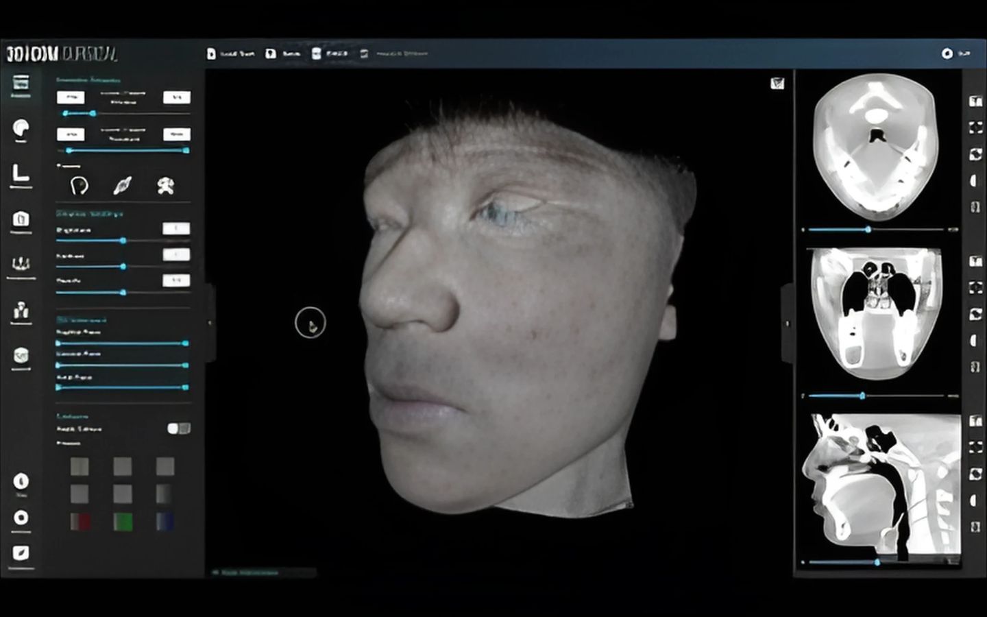
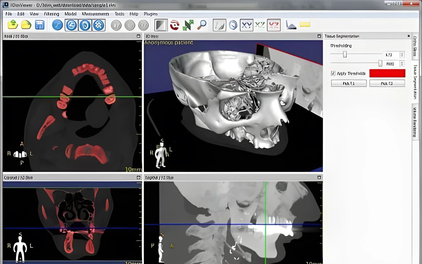
6. 3-D Volume Rendering (VR)
The 3-D VR (extent rendering) device lets you visualize huge volumes of statistics generated by way of present-day CT/MR scanners in three-dimensional space. The distinct factors of the information set may be interactively explored in the 3-d VR window.
This device helps you to rotate the extent, exchange zoom levels, and function.
a) Time-depth curves
DICOM Viewer lets you visualize the lesions’ enhancement conduct (e.G. In Breast MRI) by means of plotting time-intensity curves (TICs).
Different kinds of curves may be received: Ia – instantly (the sign depth maintains to growth over the complete dynamic length) / Ib – curved (the time-signal intensity curve is flattened within the past due postcontrast period), II – plateau (the sign depth plateaus within the intermediate and overdue postcontrast durations) or III – washout (the sign depth decreases (washes out) in the intermediate postcontrast duration).
b) Support for multitouch capabilities on Windows 10
Multi-contact aid
If you have a Windows eight or Windows 10 contact-enabled tool, you would possibly find that gestures (motions that you make with one, two, or more palms) are simpler to use than a mouse or keyboard. RadiAnt DICOM Viewer allows users to utilize the array of multi-touch gestures:
Touch the picture with one finger and pass it to browse via pix of the displayed series.
To zoom in or out, touch two points on the picture, after which move your hands far from or toward each other. Drag the image with two hands to transport it and show the invisible components of the zoomed picture.
You can change the window settings (brightness/contrast) by way of touching the image with 3 fingers and moving them up/down (brightness) or left/proper (contrast).