
Radiology: What Is It?
Radiology is a group of tests that take photos or images of the body. It is also known as diagnostic imaging. The topic covers two fields that apply radiant energy for finding and treating disease: diagnostic radiology and radiation therapy. X-rays, MRIs, ultrasounds, CT scans, and PET scans are among the most used imaging tests, though there are a few unique ones.
A radiologist will observe the final results of a certain imaging check to discover an applicable image that evaluates and makes a diagnosis. These people are normally medical doctors (MDs) with exceptionally specialised schooling targeted on the interpretation of clinical imaging.
Radiologic technologists are also useful resources in this procedure, as they use and manipulate the machines within the process of manufacturing an image.
When it comes to healthcare, early and accurate diagnosis is prime. At ALM Viewer in Des Plaines, IL, we recognize how essential it is to get the right information fast. That’s where Radiant Radiology is available in — a modern and green way to view and apprehend diagnostic photographs.
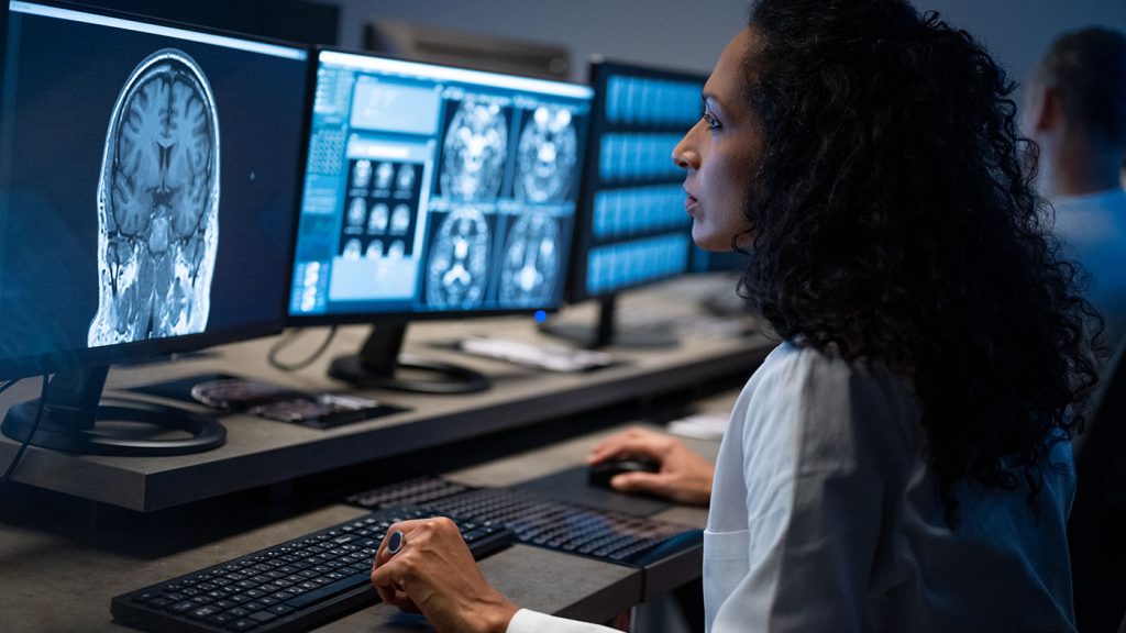
What Is Radiant Radiology?
Radiant Radiology isn’t a new kind of experiment or take a look at. Instead, it refers to the advanced software program that lets doctors and radiologists view, analyze, and percentage scientific images together with X-rays, MRIs, CT scans, and ultrasounds — all in one location.
Think of it like an upgraded model of a photograph viewer, but designed specially for scientific snapshots.
At ALM Viewer, we use the Radiant Radiology software program to make the photograph viewing process quicker, clearer, and greener.
History of Radiant Radiology
Radiant radiology started out in Germany in 1895, whilst Wilhelm Conrad Röntgen made an energized, lightproof cathode ray tube that began to fluoresce whilst situated multiple feet faraway from a screen painted with fluorescent fabric.
He knew the display screen was responding to unknown rays transmitted at some point in the room, which he had known as “x-rays.” After Röntgen’s discovery, human beings commenced creating radiography images that began as an irradiation burst and produced an opposite image on film.
Why Does Radiant Radiology Matter?
Here’s why Radiant Radiology viewer makes a difference for patients and healthcare providers:
- Faster Results
Doctors can see your photos properly, main to quicker diagnoses and treatment plans.
- Better Image Quality
High-decision pix imply clearer details, helping radiologists make greater accurate assessments.
- Easier Access
With the entirety in one system of radiant radiology, there’s no need for CDs, USBs, or printed movies. Your pics can be accessed securely through your health practitioner — every time, anywhere.
- Smart Tools
Radiant Radiology comes with clever gear that assists with degree, zoom, and examine pics without problems.
Why Is Radiant Radiology Important?
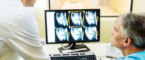
Doctor Showing Patient Radiant Radiology X-Ray
Every sector in the health care area is based on radiant radiology, which includes:
- Surgery
- Pediatrics
- Obstetrics
- Oncology
- Trauma-response
- Emergency medicinal drug
- Infectious sickness
Correct diagnosis can save lives in numerous situations, particularly for cancer patients. Family physicians and emergency rooms rely on radiology to determine the appropriate analysis and course of treatment because they are unable to manage patients without diagnostic imaging.
What is Radiant Radiology Used for?
Depending on the type of radiant radiology and any particular scan employed, radiology can be utilized for an array of illnesses. The many imaging tests include:
- Radiography: X-rays used for exploring the stomach, chest, or bones are called radiography.
- CT (Computed Tomography): A CT captures more than one x-ray angle of the patient the use of a doughnut-shaped device, then creates laptop-processed images.
- MRI (Magnetic Resonance Imaging): An MRI makes use of magnetic fields and radio waves with computer processing to create pictures.
- Mammograms: Specialized X-rays that look at breast tissues.
- Ultrasound: An ultrasound uses sound waves to create transferring snap shots that display on a monitor, normally used for echocardiograms and inspecting the womb throughout pregnancy.
- Fluoroscopy: X-rays that give real-time, moving pictures of the body. This imaging is important for lots of approaches, in particular the ones involving the gastrointestinal tract.
- Nuclear medicinal drug: These are brief-appearing radioactive substances that generate mild from bodily tissues. A digicam collects the mild, so a pc can system it and create a photo.
Diagnostic radiology makes use of these imaging effects to identify an extensive range of problems, from broken bones to coronary heart conditions and blood clots. Interventional radiology additionally makes use of imaging, along with CT scans, MRI, and ultrasounds to guide scientific tactics.
Patients are commonly awake throughout these tactics, whether it’s treating most cancers, back pain, or liver and kidney problems. In a few instances, interventional radiology eliminates the need for surgery and scopes.
Benefits of Imaging Using Radiant Radiation
Benefits of Medical Imaging
There are many blessings for patients from clinical imaging. Images of the human frame are created using quite a few methods consisting including ultrasound, magnetic resonance, nuclear medicine, and X-rays, to allow physicians to look within the frame in radiant radiology, to become aware of and/or rule out medical issues, and to diagnose illnesses. Much has lately been written approximately radiation, so it’s far vital to have some information about imaging done using radiation, especially the benefits to the affected person, at the side of the associated dangers.
Keep in mind that the radiation doses used in clinical radiological imaging examinations like computed tomography (CT) and X-ray scans are a great deal decrease than those used in radiation oncology, which makes use of radiation as a remedy to deal with most cancers. Radiant Radiology (imaging) and radiotherapy (cancer remedy) are quite different.
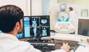
CT Benefits in Radiant Radiology
A patient struggling with a right brain stroke as proven on CT, CT perfusion, and CT Angiography.
The present-day use of imaging has been one of the wonderful advances made within the practice of medicine, allowing doctors to diagnose and control their patients’ sicknesses appropriately and swiftly. Many of these days’ medical enhancements require exams that use radiation to verify diagnoses, plan management, and reveal the reaction to treatment.
Whether it’s a basic chest X-ray for a cough, a bone X-ray for a broken leg, or something more complex like a CT test for imaging has helped a great number of people in the United States. Dr. Wilhelm Roentgen won the first Nobel Prize in Science in 1895 for producing X-rays.
CT ranks as one of the pinnacle five scientific traits in the closing forty years, consistent with most medical surveys. It has proven to be so valuable as a clinical diagnostic device that the 1979 Nobel Prize in Medicine was awarded to the inventors of CT.
CT enables more powerful clinical management, and its blessings encompass:
- Determining whether surgical procedures are necessary
- Reducing the need for exploratory surgeries
- Improving cancer prognosis and treatment
- Reducing the length of hospitalizations
- Guiding treatment of common situations such as harm, cardiac disease, and stroke
- Improving affected person placement into suitable areas of care, which include an in-depth care unit
Early Uses and Developments
The discovery of X-rays captured the clinical network’s and trendy public’s attention and creativity. This novel, invisible form of radiation, capable of penetrating strong items and revealing their inner shape, has become seen as a wonder of the modern age.
The speedy dissemination of Roentgen’s findings, facilitated with the aid of the current invention of the telegraph and a more and more connected global medical community, brought about a flurry of experimentation and alertness internationally.
Immediate Interest in X-rays
The doctor’s tests were immediately duplicated by professionals and physicians around the globe. There was an experience of exhilaration approximately the capacity applications of this new form of radiation.
Private labs, hospitals, and universities started investigating the characteristics of the X-ray view box. There was a hurry to recognize and harness this new era, regularly with little regard for protection, as the dangerous consequences of radiation were not yet understood.
Early Uses in Medicine
One of the earliest and maximum significant applications of radiant radiology of X-rays was in the field of drugs. Physicians started using X-rays to detect and treat a variety of clinical illnesses within weeks following Roentgen’s claim.
With this incident, X-rays were utilized for clinical testing for the first time.
Soon after, X-rays in radiology were utilized to find bone fractures and shots. They have come to be a critical device in orthopaedics and surgical procedure, allowing doctors to peer inner a patient’s body without the need for invasive techniques.
The radiation was used in the forces before the Great War
At the onset of World War I, the huge use of X-rays type of radiology in the army context became apparent. X-ray equipment was set up in field hospitals to aid in the care of sick soldiers.
These X-ray gadgets performed an essential function inside the conflict, considerably enhancing the treatment of gunshot and shrapnel injuries. Surgeons ought to locate and dispose of projectiles as they must, reducing contamination fees and enhancing restoration instances.
Future of X-ray Technology
The destiny of X-ray generation guarantees similar innovations and advanced radiology pushed by means of ongoing research and technological trends. The goal of these future directions is to improve the capabilities of present-day radiant radiology, is X-ray packages, and discover new areas in scientific therapy, diagnostics, and history.
Future Developments in X-ray Technology
- Higher Resolution Imaging: Research is ongoing to broaden X-ray imaging strategies that can provide even higher-resolution snapshots. This development could allow for more unique and particular visualisations of inner systems, potentially at a mobile or molecular level, substantially helping in early ailment detection and diagnosis.
- Reduced Radiation Exposure: A key location of cognizance is the reduction of radiation exposure to patients at some stage in X-ray techniques. Advancements in detector technology and photo processing algorithms are expected to yield extra efficient systems that require less radiation to supply extraordinary pics.
- 3D Imagery and Holographic imaging: There has been a rising interest in 3D X-ray technology and holographic imaging. This technology should offer three-dimensional perspectives of internal systems without the need for invasive processes, providing an extra complete diagnostic device.
- Portable and Affordable X-ray Devices: The development of smaller, extra transportable, and low-power X-ray gadgets ought to make this generation greater accessible, especially in remote or under-resourced regions. This would facilitate wider use in loads of settings, from small clinics to catastrophe zones.
Modern Applications
The X-ray era is the backbone of clinical imaging today, radiant radiology, providing vital details about the human body for both medical and treatment purposes. Its packages reflect its wide range and critical nature by reaching far outside the field of medicine into the worlds of business and science.
Modern Applications in Medical Imaging
- Radiography:
The most common use of radiation is in radiography, which refers to the most commonly employed X-ray imaging tool for viewing the internal organs of the body, especially the bones. It is commonly utilized to identify conditions and fractures, as well as and on a particular occasion, it is used to identify lung and heart diseases.
- Scans with CT, also computed tomography:
CT scans constitute a big improvement in the X-ray era. CT scans create cross-sectional images (slices) of bones, blood vessels, and clean tissues in the body with the aid of taking multiple X-ray pictures from one of a kind angles and using computer processing. This offers greater precise data than regular X-ray snapshots, beneficial in diagnosing cancers, cardiovascular illnesses, infectious illnesses, trauma, and musculoskeletal problems.
- Fluoroscopy:
Fluoroscopy includes using X-rays to gather actual-time transferring photographs of the internal structures of a patient. This approach is especially beneficial in the direction of diagnostic and therapeutic procedures, which consist of catheter insertions, angiographies, and orthopaedic surgical techniques, bearing in mind live guidance of those techniques.
Industrial Applications
- Non-Destructive Testing (NDT):
X-rays are a type of radiant radiology that is widely used in businesses to check both structures and materials non-destructively. This program is essential for identifying faults or cracks in pipelines, joints, and metallurgical procedures, especially within the production, automotive, and military industries.
- Quality Control:
X-rays are used in manufacturing to manipulate things in a pleasant way, verifying their safety and integrity. This is especially true in the electronics business, where they are used to examine circuit boards, and in the food industry to find hidden electronics in food packaging.
Astronomical Applications
- Astronomy and Astrophysics: In the field of astronomy, X-ray telescopes are used to examine and examine X-ray emissions from celestial bodies together including black holes, neutron stars, and supernova remnants. X-rays emitted through these high-energy and excessive environments in space offer precious facts about the properties and behaviours of these astronomical items.