
Medical imaging makes use of a variety of technologies to gain a deeper understanding of clinical troubles and attain a correct analysis. Let’s examine the two most important pieces of technology in this field, DICOM (digital imaging and communication in medicine) and PACS (Picture Archiving and Communication System), which provide a better understanding of devices and processes used for image modification and analysis.
In addition to checking its packaging outside advanced radiology, the purpose of this article is to clarify the difference between DICOM and PACS, their functions in clinical imaging, and their inter-personal.
What is DICOM?
DICOM means digital imaging and communication in the medical field. It is a worldwide trend used for storing, transmitting, and copying scientific imaging information and associated records.
Importantly, DICOM is not a special piece of hardware or even software. Rather, it’s an expert widely used to assist in manipulating the processing of digital images within the scientific field.

History and Development of DICOM
The National Electrical Manufacturers Association (NEMA) and the American College of Radiology (ACR) collaborated to create DICOM in the early 1980s. The goal is to standardize the conversation of medical imaging facts to improve interoperability amongst unique systems and gadgets.
Before DICOM, every piece of medical system required a proprietary device or software to examine and translate the pixels produced into something beneficial. DICOM makes it simpler for hospitals and other healthcare centers to apply equipment from particular producers while not having to pay for delivery.
Key Features of DICOM
Medical Imaging Standardisation: DICOM guarantees that photos and related statistics from specialised equipment and manufacturers are suitable.
File Format and Communication Protocols: DICOM specifies each the layout of the clinical photographs and the protocol for transmitting them.
Common Uses Of DICOM:
DICOM is used in numerous medical fields, which include:
Radiology: For X-rays, CT, MRI, and scientific imaging opinions.
Cardiology: For imaging strategies, inclusive of echocardiogram and cardiac MRI.
Additional medical requirements: In fields like dentistry, pathology, and dermatology, imaging is vital for diagnosis and remedy.
Any piece of a scientific system that produces a photo of a few type should follow the DICOM requirements. Since DICOM is also a report format, scientific imaging devices need to all produce DICOM pix that may be accessed and viewed by way of different system that meets DICOM standards.
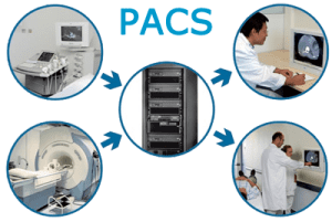
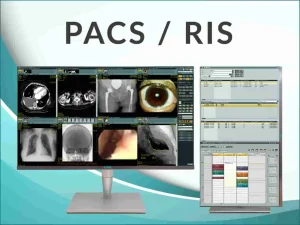
What is PACS?
PACS stands for Picture Archiving and Communication System. It is a scientific imaging generation used for storing, retrieving, managing, and dispensing scientific snapshots.
PACS doesn’t confer with a document format or photo format, but as an alternative to the way the images are saved, accessed, and transmitted among doctors and affected persons, in-house experts and external experts, and more.
History and Development of PACS
PACS was developed as a strategy to address the inefficiencies of traditional movie-primarily based imaging, permitting virtual storage and smooth get right of entry to to images. Today, PACS consists of on-web page and cloud-based solutions, and can be customized depending on the facility.
Key Features of PACS
Storage, Retrieval, Management, and Distribution: Picture Archiving and Communication System PACS handles the complete lifecycle of clinical photos from acquisition to archiving.
Integration with Hospital Information Systems: PACS frequently integrates with other structures like Electronic Health Records (EHR) for comprehensive patient care.
Common Uses of PACS
Although radiology is the primary software for PACS, its programs additionally consist of:
Radiology: For controlling CT scans, MRIs, X-rays, and other tests.
Other Medical Fields: Such as oncology and orthopedics, in which imaging is important.
DICOM and PACS: Understanding the Relationship
The relationship between PACS and DICOM is symbiotic. PACS is predicated on DICOM to ensure seamless communication, at the same time as DICOM provides the framework for PACS to control images efficiently.
Key Differences
System vs. Standard: PACS is a comprehensive machine for dealing with scientific images, whilst DICOM is a popular protocol for communication.
Functionality: PACS affords functionalities for the garage, retrieval, and manipulation of photos, whereas DICOM ensures that those photographs can be exchanged and interpreted efficaciously.
Implementation: PACS involves the deployment of hardware and software solutions, even as DICOM includes adhering to precise communication and statistics formatting requirements.
How They Work Together
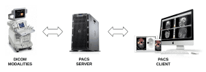
DICOM and PACS work collectively to create a cohesive scientific imaging environment. When an image is received from a modality (e.g., MRI scanner), it is formatted in keeping with the DICOM standard. This DICOM photograph is then sent to the PACS, wherein it’s further saved, managed, and distributed to healthcare vendors.
Consider this practical example: A radiologist perspectives a CT test on a PACS laptop. The CT experiment has acquired the usage of DICOM standards, making sure that the image is displayed efficiently on the workstation, irrespective of the scanner’s producer. This seamless conversation is critical for accurate diagnoses and well-timed interventions.
Practical Applications in Healthcare
The realistic applications of DICOM and PACS extend to diverse elements of healthcare, improving each medical and technical workflow.
Clinical Implementation
In a medical institution, putting PACS streamlines the picture control technique, permitting healthcare vendors to get access to photos fast and correctly. This ends in faster diagnoses, reduced wait times, and advanced patient outcomes.
Benefits for healthcare carriers:
Improved get entry to to pictures: Healthcare vendors can view snapshots from any location within the community.
Enhanced collaboration: Multiple professionals can evaluate photographs simultaneously, facilitating higher decision-making.
Reduced errors: Digital photos get rid of the threat of misplaced or broken film.
Technical Considerations
Implementing DICOM and PACS includes several technical considerations, along with gadget requirements, integration demanding situations, and best practices. Healthcare centers need to ensure that their systems observe DICOM requirements to maintain interoperability.
What Separates DICOM and PACS?
Fundamental Differences Between DICOM and PACS
DICOM is a technical format for a photo, in addition to a report layout for photographs. The tool used to get right of entry to the pics is known as PACS.
DICOM: A communication and formatting popular for images.
PACS: A machine for archiving and managing photographs.
Or to put it an exceptional way, a DICOM photograph will be accessed via a health center’s PACS device.
How DICOM and PACS Complement Each Other
DICOM and PACS work collectively. Since DICOM is the technical standard, the majority of radiology photos handy in a medical facility’s PACS have to be within the DICOM format.
DICOM: Ensures that images are successfully formatted and transmitted among devices.
PACS: Provides the infrastructure for storing and copying those photographs effectively.
How is DICOM Used with PACS?
Role of DICOM and PACS
DICOM is essential for making sure that snapshots from diverse imaging devices can be stored and accessed within PACS radiology. It standardizes the photo codecs, permitting seamless integration.
Workflow Example: From Image Acquisition to Storage and Retrieval
Image Capture: Medical snapshots are acquired the use of imaging devices.
DICOM Formatting: These pictures are formatted in step with DICOM requirements.
PACS Storage and Retrieval: Formatted photographs are stored in PACS and may be retrieved as needed.
Can PACS Work Without DICOM?
Theoretically Possible Scenarios
PACS ought to probably use proprietary formats and communication protocols. A PACS isn’t constrained to the DICOM format, but because DICOM is the widespread trendy for medical imaging equipment, almost all PACS will recognition on processing and managing DICOM documents.
Practical Limitations and Challenges
Lack of Interoperability: Without DICOM, specific imaging gadgets and structures might not be compatible.
Integration Issues: Non-standardized records can cause big challenges in integrating with other healthcare systems.
Advantages of Using DICOM and PACS
Improved Interoperability: Ensures distinct systems can work together seamlessly.
Medical gadget producers and each person providing a PACS solution normally focus on DICOM, especially with the medical industry’s consciousness of the management and protection of records, which are made simpler with a unified system.
DICOM and PACS Advantages in These Fields
Enhanced Image Management: Streamlines the procedure of storing, retrieving, and sharing scientific snapshots.
Improved Patient Care: Facilitates well-timed and accurate prognosis and remedy planning.
Even past radiology, DICOM and PACS work together to improve the ease of dealing with medical images and supply health-related results quickly.
What are the traits of DICOM and PACS?
In modern clinical imaging, DICOM and PACS are critical equipment that allow healthcare companies to handle medical images effectively and efficiently. They are important equipment for diagnosing and treating clinical situations, supplying correct and well-timed facts to healthcare experts.
How does DICOM and PACS work?
To manage medical images, DICOM and PACS, two related technologies, combine. Here’s how they work:
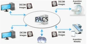
Image Acquisition: Images are taken and saved in DICOM format through medical imaging gadgets, including MRI scanners and X-ray machines. Information concerning the patient, imaging equipment, and image is all contained in the DICOM format.
Image storage: PACS, which acts as a central repository for images, is the place where the DICOM image is then placed.
Viewing image: Healthcare professionals, DICOM viewers can see DICOM images using special software. These audiences allow them to manipulate and analyze images for accurate diagnosis and treatment decision-making.
Image communication: DICOM and PACS use communication protocols to facilitate the transfer of images between various healthcare systems. The protocol guarantees that photos are sent to a uniform format, allowing them to be seen by various computers without any incompatibility.
Difference Between DICOM and PACS
Although each DICOM and PACS is are important technology within the discipline of clinical imaging, their features range.
Along with X-rays, CT scans, and MRI snapshots, it’s a general for the trade and handling of scientific imaging records. DICOM specifies how medical snapshots must be received, stored, transmitted, and displayed. DICOM allows the change of scientific images and associated records between exceptional imaging systems, hospitals, clinics, and different healthcare businesses. To put it another way, DICOM is well-known for the photographs’ layout, communication strategies, and accompanying metadata.
Picture Archiving and Communication System is another name for PACS. It is a clinical imaging generation that is used to AR keep, retrieve, distribute, and show medical photographs, together with X-rays, CT scans, and MRI images. It allows healthcare specialists to access and think about images from any area with net get admission to. A photo acquisition device, a database or image archive, and a display or viewing station make up a PACS.
DICOM Features
DICOM (Digital Imaging and Communications in Medicine) is a standard for the communication and control of clinical imaging data, and it consists of several capabilities that enable interoperability among distinct scientific imaging devices and software programs. Here are some of the important capabilities of DICOM:
Standardized Image Format: DICOM defines a standardized image layout for scientific photographs, which guarantees that snapshots may be regarded and interpreted on one of a kind imaging gadgets and software applications.
Metadata: A standardised collection of statistics approximately the image, or metadata, is protected with DICOM. Patient demographics, imaging modalities, acquisition parameters, and image comments are the various information included in this metadata. Because the metadata is stored in a uniform format, inexperienced users and changes to snapshots and associated facts are possible.
Image Compression: DICOM consists of numerous picture compression techniques that allow the effective transmission and storage of photographs without sacrificing image excellent.
Network Protocols: To transmit scientific images and related files, DICOM specifies a whole lot of community protocols. These protocols make sure that photographs can be transmitted securely and successfully over various types of networks, together with the internet.
Security and Privacy: DICOM consists of functions for protection and privacy, consisting of encryption and authentication, which ensure that scientific images and associated data are transmitted and saved securely.
Workflow Management: DICOM defines workflows for the acquisition, storage, retrieval, and display of scientific snapshots and related information. These workflows enable green management of scientific photos and associated facts across exceptional healthcare organizations and imaging devices.
PACS Features
A software program called PACS (Picture Archiving Communication System) is used in clinical facilities to save, retrieve, and disseminate medical photographs and the related patient statistics. Some of the important functions of PACS include:
Image Storage: PACS gives a centralized digital archive for storing clinical photographs, allowing healthcare companies to access and review affected person photographs from anywhere in the facility.
Image Retrieval: PACS permits healthcare carriers to retrieve affected person images quickly and effortlessly, eliminating the need to manually search through bodily image documents.
Image Distribution: PACS allows the sharing of medical images and related data among healthcare carriers and centers, improving collaboration and facilitating second reviews.
Image Viewing: PACS affords numerous tools for viewing and manipulating scientific images, such as zooming, panning, and changing assessment stages, to enhance visualization and useful resource prognosis.
Security and Access Control: PACS has integrated protection capabilities to ensure the affected person privateness and shield sensitive clinical statistics. Access to patient pics and records is confined to legal healthcare carriers.
Integration with Other Systems: PACS may be integrated with different healthcare statistics structures, along with digital health statistics (EHRs) and radiology information systems (RIS), to offer an entire picture of the affected person’s fitness records.
Conclusion about DICOM and PACS
For medical professionals involved in clinical imaging, it is necessary to understand the difference between DICOM and PACS. While DICOM standardizes the layout and transmission of scientific snapshots, PACS affords a complete solution for storing and managing these images.
Together, they beautify the performance and effectiveness of clinical imaging, in the long run enhancing patient care. As technology advances, the integration and application of DICOM and PACS—especially through platforms like ALM Viewer—are likely to expand, further revolutionizing the field of clinical imaging.