

Overview of the DICOM standard
DICOM provides detailed engineering information that can be used in interface specifications to enable network connections between the products to a variety of suppliers. The standard specifies how to format and exchange medical images and associated information in the hospital and also (e.g., counteriaology, telemedicine). DICOM interfaces are available for connecting any combination of lower categories of digital imaging equipment: Image collection tool (e.g., computed tomography, magnetic resonance imaging, calculated radiography, ultrasonography, and core therapy scanner); image archive; image processing tools and workplace for image performance; hard-copy output unit (e.g., photographic transparency film and paper printer). These standardized connections play a critical role in Radiology Informatics, ensuring smooth interoperability across different imaging systems.
DICOM is a message standard (i.e., a specification for the difference between information between the computer systems). DICOM is a comprehensive specification of information material, composition, coding, and communication protocols for the electronic exchange of information related to clinical and therapeutic images. Some other health data exchange standards specify only a subset of the properties that affect interoperability. Health Level Seven (HL7) Standard 2 specifies a message model, but provides only a brief network communication specification, whereas Radiology Informatics leverages both DICOM and HL7 to ensure seamless integration and data flow across imaging systems.
DICOM Protocols, Services, and Goods
DICOM specifies a protocol for message exchange. The free DICOM viewer message protocol provides communications frames for DICOM services. DICOM protocol is compatible with the Transmission Control Protocol and the Internet Protocol. This DICOM application allows institutions to communicate on the Internet, playing an essential role in Radiology Informatics by enabling seamless data exchange and interoperability across healthcare systems.

This figure refers to three separate implementations of the DICOM Protocol scheme. A DICOM network connection is present between the application program (Peer Dicom application institutions) of imaging equipment. Application software in Configuration 1 produces DICOM Command requests (RQ) and command responses (RSP) messages flowing from one device to another. A separate DICOM message service element (DIMSE) in configuration 2 produces command messages from the machine application software. The DICOM Protocol machine is the DICOM supplier (DSP), which plays a key role in Radiology Informatics by enabling seamless communication and interoperability between imaging systems.
Application software is DICOM Service User (DSU). Configuration 3 uses separate modules for all communication and application features. A second layer of free DICOM viewer messages is used in each device. This is DIMS Service Primters: Request Primitive (Recup), Indication Primitive (INPP), Response Primitive (RSPP), and Confirmation Primitive (CFMP). A standalone DSU module app produces DIMSE Service Primters from the application software. Although the protocol and user modules of the DICOM service can be used in different ways, the external command messages are the same for all configurations.
DICOM services come in two groups: total and generalized. The total services were designed for compatibility with previous versions of the ACR NEMA standard. They had originally intended for storage (C-store), Querry (C-Find), recovery (C-street), and image transfer (C-Move). However, overall services are also useful for other types of information, such as interpretation reports. Note that the total group does not include the “update” service, which is an important aspect in Radiology Informatics for ensuring smooth data exchange and workflow efficiency.
This lapse is conscious. Architects of the original ACRnema standard chose to publish “updates” to reduce the possibility of changing an image mail. Thus, overall services are adapted to the exchange of images. However, this adaptation limits the use of overall services for other domains. Explanation: Data exchange is an area where total services are useful. Since the change of medical records is absolutely prohibited, changes to original interpretation reports are usually published as new documents. This business model is translated accurately to the total service paradigm, which remains a key principle in Radiology Informatics for maintaining data integrity and compliance.
Generalized services were designed to provide extensive information management functionality. Note that the name “generalized” is not related to the normalization of the database. Normalized services were intended for use with items representing the properties of a world unit, while overall services were originally used with documents (images) containing information from more than one real-world unit (eg, pixel data, equipment, and patient identification number). Generalized services support basic information management operations: N-CREATE, N-DELETE (N-DELETT), Update, and Pickup (N-GET). In addition, domain-specific operations such as “Print a Sheet of Film” can be defined. An alert service is also specified in the generalized group, highlighting its importance in Radiology Informatics for managing complex imaging data workflows.
Despite the flexibility, the generalized service group has some remarkable limits. “Sequence to the item” in the Update Service (N-set) has limited data type tools. The N-set must update an entire sequence instead of a personal information element in a sequence. The generalized group also lacks a query service. This flashing lapse is the result of a lack of industry in the network question protocol at the time when the standard was written, which created additional complexities later addressed in Radiology Informatics advancements.
For the ISIS interface for information system implication system (ISIS), this limit is modified by the original model’s work object pairs (see the definition of the SOP class in the next section). The modeling worker specifies a composite query service to restore demographic and plan information from SOP Class Imaging Equipment, which highlights the role of Radiology Informatics in improving interoperability and structured data management.
The real-world institutions (eg, images, processes, or interpretation reports) are represented in the DICOM Sentic data model with templates for properties. The formal specifications of these templates are documented in the DICOM information object description (IODs). An IOD is an abstract description of a square of institutions. An order given by values representing the characteristics of a member of a class can be served by one or more DICOM overall or generalized services.
A DICOM Service-Object-Page (SOP) class specifies a combination of an IOD and a set of services (Dimse Service Group) that are useful for a given purpose. SOP classes (such as the Basic Modelity Works SOP class) are specified in the service classes according to their purpose. SOP classes that use total services are composite. Generalized SOP classes use generalized services. An example of the SOP class is known as an example of a service-object-Zodi (SOP). The overall object and generalized objects are synonymous for general and generalized SOP examples, making them an essential concept in Radiology Informatics for standardizing imaging workflows.
DICOM object vocabulary is generous, but it is definitely accurate. For brevity, DICOM SOP classes are often referred to as objects or information objects. Note, however, that DICOM objects are “static” objects. They are passive information structures that can be operated in external ways. They are not independent software components capable of polymorphism, encapsulation, and inheritance. Their design is in line with their purpose. DICOM SOP classes (and examples) are useful for data exchange. They are not application components. The DICOM SOP class data structure maps the DICOM Service groups for the data structures of software components and object methods, which is a critical aspect of Radiology Informatics in ensuring interoperability and standardization across imaging systems.
The Message Transaction Association begins with installation using DICOM. A DICOM Association is a communication session consisting of a couple of colleagues’ DIMS service users (see Img1). In other words, a DICOM Association is an open channel for message exchange between two devices using the DICOM Protocol Machine (software) and produces DICOM messages. This structured communication process plays a vital role in Radiology Informatics, enabling seamless data exchange and interoperability between imaging systems.
During the installation process of the association, two tools come to a shared understanding of information structures, which will be exchanged, and services will be implemented (ie, Essence Syntax). Further parameters required for interoperability, such as the City Creation and Data Compacting Method, are also involved (ie, transmission syntax). The association is known as the Association Control Service Element (ACSE). The DICOM Protocol specifies coordination of ACSE and DIMSE functionality, which is a critical foundation for Radiology Informatics in ensuring smooth communication and interoperability across imaging systems.
DICOM service classes support five general application areas. Each will be described in separate sections as follows. Enable service classes:
1: Network image administration
2: Network image Interpretation management
3: Network Print Administration
4: Imaging process management
5: Off-line Storage Media Management
1: Network image administration
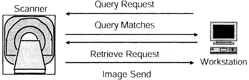
DICOM Network Image Management supports two common references on interaction between imaging units: Push mode and Pull mode. The original service is “Push” mode, where one device only sends images to another device on a computer network (IMG. 2). “Bridge Mode” is a more elaborate two-stage process that allows the user to first ask for an external device and then rebuild selected images (IMG. 3). These modes play a vital role in Radiology Informatics, ensuring efficient image transfer and accessibility across different systems.
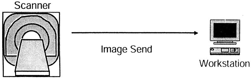
Image transfer. The scanner begins regular image transfer. A DICOM scanner does not specify the behavior of the device; Each time it is ready, scans can start sending pictures. This can be done automatically because individual images are completed during a scanning process, or it can be done after some time when all images of a procedure are obtained and the scanner operator has started transfer by activating the “Image” key on the scanner console. Such image-sharing processes are fundamental to Radiology Informatics, as they ensure smooth communication and accessibility of medical imaging data across systems.
When the scanner is ready to send, it sends pictures to the workstation one by one. The scanner initiates a DICOM communication session (called “Association”) with a workstation for the transfer of each image. Different details are interacted with during the installation of the association, so that the workstation can prepare to handle the image that is about to be achieved. This seamless transfer process highlights the importance of Radiology Informatics in ensuring accurate, efficient, and standardized image exchange between systems.
2: Networking Image Interpretation Management
DICOM Standard Supplement 23 is a network image interpretation object (structured interpretation SOP class), and a related interpretation defines a storage service. At this time, the specification is under a formal modification control process, and it will be frozen for testing implementation. This advancement plays a crucial role in Radiology Informatics, supporting structured reporting and standardized interpretation across medical imaging systems.
The structured interpretation information uses SOP Class Specification General Service Group. Therefore, the same set of services is used for images for structured interpretation objects. A structured interpretation SOP example expresses comments that form part of the results of an imaging process. The complement shows the set of 23 observation spaces defined for observation reporting in DICOM. The comments can be linked to other comments through the relational types specified.
Complement 23 texts, codes, and the ability to connect the measurement concepts to the coordinated sets (i.e., connecting comments for image features that develop observer decisions). This capacity is beyond the possession of the comments. This supervisor enables documentation of knowledge, which is an essential part of Radiology Informatics for enhancing structured reporting and clinical decision-making.
3: Network Print Administration
DICOM Network Print Management enables procurement tools and workstations to share a printer on the DICOM network (IMG 5), as individual computers share a network laser printer. Appendix H of PS 3.4 specifies the pressure management class. 2 DICOM Print Management Specification defines a Main set of compulsory features and some alternative extensions, supporting streamlined printing workflows in Radiology Informatics for diagnostic imaging environments.
Four Meta SOP classes (seen with inter-oriented items used in a coordinated manner) are specified to support basic print applications. Mandatory meta SOP classes support (1) Basic grass scale printing, (2) Basic color print, (3) Grasscale print with lookup table improvement, and (4) color printing with 4 lookup table improvement. At least one of the four compulsory meta SOP sections should be used to claim to be in line with the standard. Any combination of alternative SOP classes can also be used. Alternative SOP classes enable increased reporting of film anti-cantile, image overlay, and print status for job performance, making these functions highly valuable in Radiology Informatics workflows.
4: Imaging process management
Study administration SOP class and study component management. The SOP class provides extensive capacity for the imaging process. Study the SOP incompost map for requested procedures, and the study component for the performance process stages on the SOP incident map. They are generalized SOP classes. For example, they support the N-set and N-event_report services and provide government management and event warning facilities. Complement 17, Performance Process Phase Management, is close to the implementation of this writing time.
This draft supplement provides a small number of new properties for the details of the supplementary imaging processes and enables better connection of the study component (executive process phase) with the new DICOM model list SOP class (see below for further details), which highlights their critical role in Radiology Informatics.
DICOM Composite Query-Retrieval Model (see above) provides a mechanism for the construction of a hierarchical database of patients, studies, chains, and images. This image model has been used extensively as the basis for the image processing processes database, especially where image collection devices are not connected to the external information system (IS). In this non-world scenario, the procedure can be assigned by the imaging equipment of the identifiers. This is another system; the process identifiers must be covered with post facto, which demonstrates its significant role in Radiology Informatics.
5: Off-line Storage Media Management
DICOM Off-Line Storage Administration allows users to exchange DICOM files on removable storage media manually. The 3.5-inch is a compact slice memory (CD-ROM) plate and a DICOM file format for the optical plate. A DICOM file can not only include images, but also related information that sets one image apart from another (eg, relevant details of the performance process, interpretation text, or format settings for printing), making it an essential component in Radiology Informatics for managing and sharing medical data.
The ability to send information related to the image is one of the most important properties limited to image data alone, and separates DICOM from multiple image resistance. DICOM specifies a file directory and a file format for pictures and associated data, making it a cornerstone of Radiology Informatics by ensuring standardized storage and exchange of medical imaging information.
To transfer DICOM data for a specific clinical imaging reference, users can designate preferred physical media types (e.g., gastrointestinal endoscopy, general diagnostic ultrasound, or coronary). Insufficient compression of general photographic experts on compact disk bus media, for instance, has been used by the cardiology community in the United States to specify coronary storage and heart catheterization photographs. Additional uses for DICOM Off-Line Storage Media Management include transferring data from a diagnostic conference to the conference room or from a diagnostic imaging unit to a medical advice firm, which highlights its practical role in Radiology Informatics for effective data sharing and collaboration.