
ALM DICOM Viewer Hybrid Model
The Future of Medical Imaging Flexibility
DICOM Viewer Hybrid Model: Cloud Efficiency Meets Desktop Performance
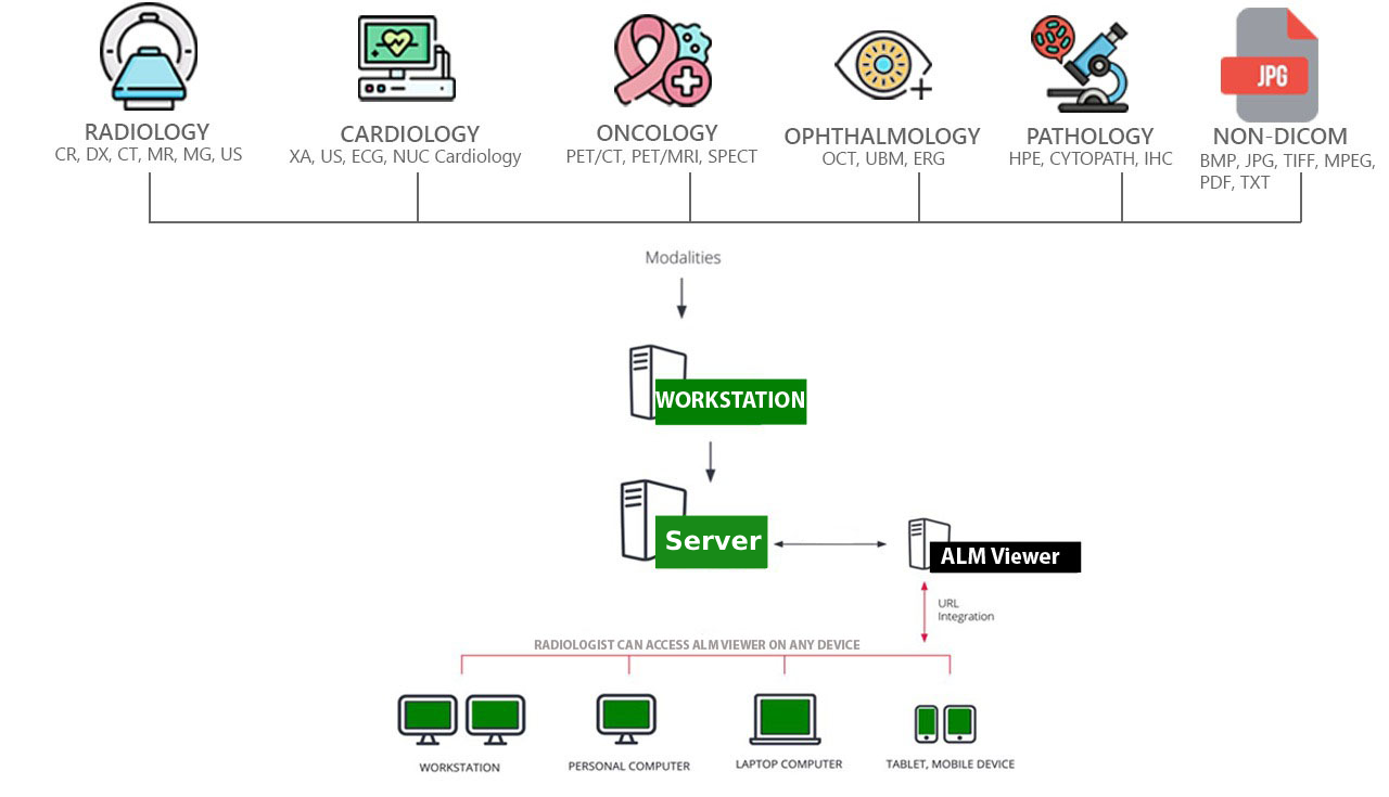
ALM DICOM Viewer Hybrid Model is a PACS viewer for medical images, designed to deliver a unique experience. With its intuitive interface and unmatched performance, It will be your go-to choice every time.
No device restrictions, you can access the Viewer on any device, any time, anywhere. Our software integrates Zero Trust Security, ensuring that every user and device is continuously authenticated and authorized. This advanced security model minimizes risks by enforcing strict access controls, providing robust protection against unauthorized access.
Local Archive
Multi-touch support
Benefits
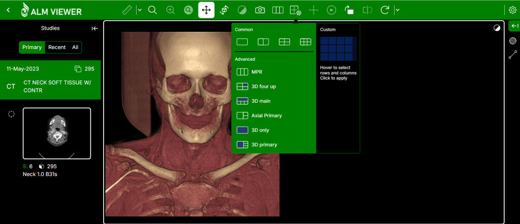
Advanced Visualization
3D – 4D reconstruction, multiplanar reconstruction (MPR), and maximum intensity projection (MIP) for comprehensive image analysis.
User-Friendly Interface
Compatibility
Annotations and Measurements
Features
Enhanced Diagnostic Accuracy
High-resolution images and advanced visualization tools facilitate accurate diagnosis and treatment planning.
Streamlined Workflow
Seamless integration with PACS (Picture Archiving and Communication System) and other medical systems optimizes workflow efficiency.
Accessibility & Collaboration
ALM DICOM viewer allow healthcare professionals to access and share medical images from anywhere, promoting collaborative care.
Cost Efficiency
Reduces the need for physical storage and maintenance, lowering operational costs.
Zero Trust Security
Ensuring that every user and device is continuously authenticated and authorized. This DICOM Viewer Hybrid Model minimizes risks by enforcing strict access controls, providing robust protection against unauthorized access from internal and external network.

Regular Audits and Monitoring

Backup and Disaster Recovery

Secure User Authentication

Real-Time Collaboration

Data Encryption

Compliance

Secure Image Sharing

Access Control
Role-based access control (RBAC) ensures that only authorized personnel can access sensitive data.
Advanced Tools for Enhanced Analysis
3D Visualization
4D Imaging
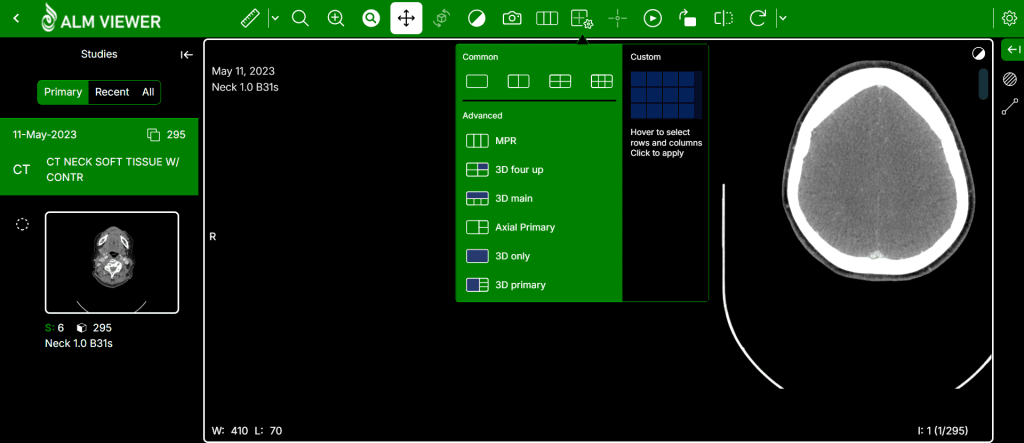
Multi-Planar Reconstruction (MPR)
Segmentation
Cine Mode
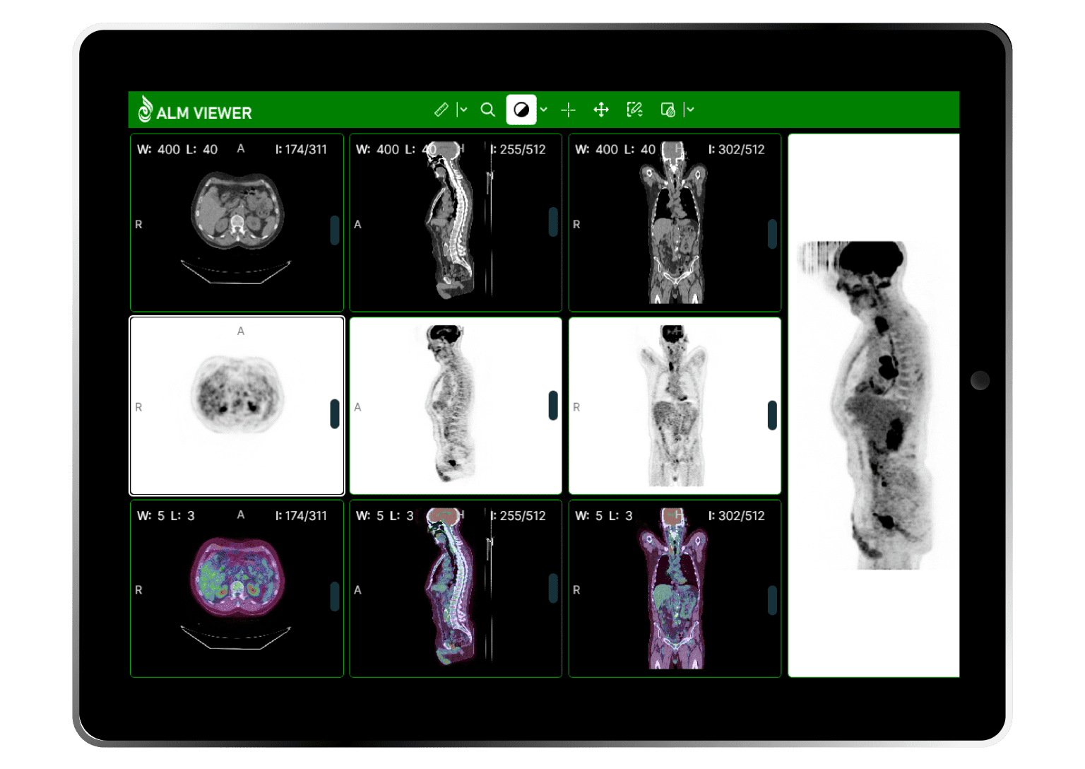
PET-CT image fusion
Overlay a color-mapped PET image onto a CT scan to obtain anatomical references for regions with increased FDG (fluorodeoxyglucose) uptake values. Use the ellipse tool to measure maximum, minimum and average values of SUVbw (Standardized Uptake Value calculated using body weight) in a specified area.
Image fusion can also be applied to other imaging modalities, such as Magnetic Resonance, e.g. DWI images can be fused with T1 or T2 weighted scans.
Essential Tools Within Reach
DICOM Viewer Hybrid Model offers a comprehensive set of basic tools for image manipulation and measurement, including:
- Smooth Zooming and Panning
- Brightness and Contrast Adjustments
- Preset Window Settings
- Image Rotation and Flipping
- Segment Length Measurement
- Parameter Analysis
- Angle Measurement
- Freehand Drawing
- Area Measurement
- Volume Measurement
- Measurement