
Challenges in Medical Imaging Data and the Role of DICOM
Diagnostic depiction fields are experiencing significant growth, after which the production of large amounts of data is increasing. Lack of standardization and privacy considerations are considered to be the main obstacles to high computerization. The purpose of this task is to confirm whether the advanced features of Apart from the DICOM standard, research techniques also make effective use of data storage imaging. The problem will be analyzed by checking the publicly shared medical imaging database and assessing how the most common medical image processing software tools, including free DICOM viewer, support DICOM in all its abilities.
Therefore, 100 public databases and ten medical imaging software tools were selected and investigated using a systematic approach. In particular, the DICOM field is related to privacy, division, and reporting is evaluated The selected database software tool is evaluated to read and write the same Dicom field. From our analysis, the Dicom-examined database uses the DICOM format to record meaningful information to manage less than one-third of the database.
When it comes to software, most people do not allow any or all DICOM regions to manage, read, and write. Surprisingly, if we inspect the sharing of the breast calculation Tomography Data to address the COVID-19 pandemic, there are only two datasets of the 12 published in the DICOM format. Our work shows how Dicom can potentially support large data management; However, more efforts are required to promote the use of existing efforts from the scientific and technical societies. standards, to encourage data sharing and differences for the solid development of large-scale data analysis.
Key points.
Standardization is important for large computer capitalization.
DICOM imaging supports the main data management functions.
Most of the shared research databases do not use the DICOM format completely.
Imaging software tools do not fully support DICOM -advanced functions.
DICOM must be fully promoted in data sharing and software development.
Modern times undergo rapid technological development, and we look at the production of large amounts of information that can be increased, qualified with the right management and analysis. We are in the so-called large data age, where the value of the data can be the real engine for innovation.
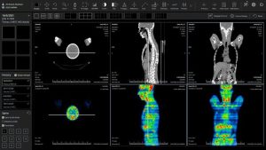
The health care system, where the information of the multi-source patient is collected regularly, faces volume and complexity. In large data types, depiction data can be considered the largest in volume. It incorporates not only gigapixel images, such as tissues or organs, but also metadata and quantitative measurements in subcontricular solutions. For example, Neuroimaging currently produces more than 10 petabytes of data each year, with a nine times increase in data complexity (i.e., data collection) over the past three decades. Tools like a free DICOM viewer are essential to efficiently access, interpret, and manage such high-volume medical imaging data.
Therefore, standardization of medical imaging formats plays an important role in the efficient utilization of data and later clinical decision-making.
WHO’s Recognition of Standardization Gaps and the Role of DICOM.
The World Health Organization (WHO) recognizes a lack of standardization, as well as the main barrier for great data utilization, with privacy considerations. It is a current standard for storing and transferring medical images of digital imaging and communication in medical format, which allows many manufacturers to work with many manufacturers with scanners, servers, workstations, printers, networking hardware products and collections and collections and communication systems (PAC). All metadata about raw imaging data, image collections, and curve procedures—including a number of processes like intelligent data, medical images, image growth, or anatomy of areas of interest in structured reporting—has been incorporated by DICOM.
In addition to DICOM, the most commonly used formats analyses are designed for the medical images format and its latest version, Neuroimaging Informatics Technology Initiative (NIFTI), Metaimage (MHD), and almost all RAW Intelligence Data (NRRD). They are largely used to store images for finishing operations. However, it is important to note that DICOM remains a standard for clinical treatment of images, which is regularly acquired in DICOM and eventually becomes other formats.
For this reason, our work does not focus on the pre-pre-consolidated use of DICOM as a clinical standard, but as a cyst bridge between the clinic and the research, on the extension of the standard towards large data analysis. Tools like the dicom radiant viewer help streamline this process by offering advanced capabilities for both clinical and research use.
In previous years, the DICOM management group has actually followed these requirements by defining and expanding the standard. Nevertheless, the simple definition of a standard is not enough to meet the needs since the availability of users and support with appropriate software tools.
This task’s objective is to assess the Dicom standard’s successful implementation from a large data perspective, specifically:
DICOM Standard: How to identify and present the current challenges in clinical imaging management and analysis.
To confirm whether the standard is used in clinical and research exercises, it is to check the public data shared by the research society and check how much the standard in the main software for the management and treatment of clinical images is really supported.
Scientific literature shows different initiatives, aimed at evaluating the use of dicom and related research goals, especially for operational imaging or de-detectives [11] for quantitative imaging for the processing of clinical data; In this work, we intend to do a comprehensive evaluation that includes all main data curing operations for both the software tool and the ongoing dataset.
The next sections elaborate on the introduction of this reference and show the current support for the DICOM format in diagnostic workflows.
Big Data -Workflow
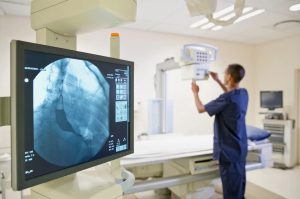
Over the past few decades, despite great progress at the beginning of the PAC and the approval of the International DICOM standard for the storage and transfer of medical imaging data, there are still significant obstacles to the effective implementation of major data analyses on diagnostic depiction data. Tools like the DICOMDIR viewer have emerged to improve accessibility and navigation through structured DICOM directories, helping bridge the gap between standardized storage and efficient data analysis.
Clinical photographs make up a significant portion of the data that is used in healthcare.
Unfortunately, medical data is often stored in qualitative reports, which do not allow for the restoration of related details along with images of interest. In fact, clinical decisions are usually made of information obtained from several sources that can vary from the patient to clinical systems, but cannot be stored in a correct and standardized way. Each of these individual properties can be regarded as a basic element of the resulting definition of an algorithm or, e.g. Monitored or unheard of model.
Confidentiality and de-identity.
One of the most talked about topics related to the collection of privacy data, analysis and interpretation is that today, these activities and their results are considered a new business. Medical data can have many legitimate secondary applications, such as research projects or teaching, but to obtain informed consent from patients is strictly necessary. Another example is the development of decision support systems, which can sufficiently commercialize the results of digital toolkits. Tools like the DICOMDIR viewer are instrumental in organizing and securely accessing anonymized imaging data, especially when managing patient records for secondary use. In addition, most individual data should be safely removed, although the link to some data or personal information should in many cases, be placed.
The increasing demand for future models, clinical decision support systems and data analysis has created a situation where research requirements often conflict with privacy rules. At this time, there is no sanctioned solution since the amount and type of individual data required by the researchers, which vary depending on the case to case, and the option for information to be kept is the option, modified or removed, depending on the goals and rules to follow it.
To overcome these problems, the authorities and institutions have developed rules and regulations that bring together privacy, data, and research goals. For this purpose, some of the most sought-after and used rules should be considered to use data for research goals:
HIPAA, 1996 Health Insurance Portability and Liability Act (HIPAA),
GDPR, General Data Protection Regulation (EU) 2016/679.
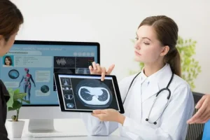
In particular, GDPR allows citizens or residents of the EU to request the deletion of their data, changes, or access, while HIPAA does not provide this right. GDPR similarly affects all personal information, while HIPAA is limited to “Protected Health Information” (PHI), which is defined as information that can be used directly or indirectly to identify a person in relation to their past, current, or future health. Tools like the radiant dicom viewer help healthcare providers manage and access such sensitive imaging data efficiently while ensuring compliance with both regulations. In addition, the HIPAA method “Safe Harbor” contains 18 identifiers for removal or modification, while GDPR has no clear list of elements that end.
There are different ways to light pseudo names and interaction. Especially ISO 25237: After the 2017 standard, there is a “procedure defined as a” process, where individual data is replaced in such a way that a data theme can be no longer directly or indirectly recognised, either by the data controller working alone or in conjunction with the other page, “data subject and one or more paps.”
From these definitions, approaches can be used according to HIPAA, and both anonymous and pseudonymous are considered “data processing” under GDPR. In the latter case, the right to access, change, and cancel the citizen’s personal data should be guaranteed. Since there is no direct or indirect link to identify the original patient with anonymous data, some additional processing or processing is made on the dataset beyond the extent of GDPR.
The implementation of detectives on clinical studies involves the discovery, elimination, and replacement of identifiers in images and tags describing them. Most DICOM elements have information on demographic and related medical images and patients, which must be confidential or removed in the event of the use of tests for research purposes. Instead of addressing cybersecurity concerns, the DICOM standard has added options for encrypting and protecting data that persists after HIPAA is implemented. network connections since 1999, as stated in the document “Security and Privacy in Dicom.” Additionally, in 2001, Dicom enhanced the use of CMS (cryptographic message syntax) to encrypt Dicom data, generating a DICOM object’s pHI encryption.
Profiles and options for removing properties in a DICOM data set and address replacement are reported in Appendix E by DICOM PS3.15 2020d. In particular, it includes a basic profile with multiple containers or pure alternatives, and other implementations such as “in standard compliance with IOD”. Each function can be replaced, cleaned or kept based on the privacy level and the importance of identity. In order to maintain the history of Metadata, the following tags must be added after the use of one of the De-determinification profiles.
DICOM images have different software tools to -identify. Aryanto et al. Provide a complete observation and important analysis.
It is very important to mention that removing or modifying the metadata in dicom images may not be enough to prevent re-identification of the subject. In fact, the patient’s information in Topogues for CT data and ultrasound images can burn the patient’s information in pixel data. To deal with this important problem, some specific studies and equipment are available to the scientific community. In addition, it is shown that people or specific software can identify individuals by reorganizing face images in cranial MRI or CT.
Further efforts are required to develop reliable de-identified methods for medical images as facial properties such as facial properties, such as facial properties. Furthermore, it is anticipated that the development of particular Dicom features that satisfy this operation would completely standardise the identification procedures.
Annotation and division
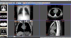
Important actions to extract basic functions from medical images for both clinical decision-making and research are annotation and classification. The annotation process focuses on labeling images with further information that is useful for the data section, classification and grouping. In particular, this routine replaces descriptive and qualitative image functions in machine-related data, and thus is suitable for automatic image analysis that monitors artificial intelligence (AI) functions.
The division can be regarded as a special case of annotation where one or more image areas are isolated in the so-called regions (ROI). Tools like the Radiant DICOM viewer support these processes by enabling precise image labeling and segmentation for advanced diagnostics and research. In fact, the partition is based on a binary mask (manual, semi, automated or completely automatic) of the pixel related to the ROI.
In the context of large data analysis, annotation and segmentation routines are basic processes for data details, sharing, and analysis. In addition, the enormous amount of the data to be administered is administered and monitored data that is capable of and is capable of standardization.
To solve the problem, the National Electrical Manufacturers Association (NEMA) DICOM-RT developed, ie, the first extension of the DICOM standard that may contain information on annotation [30]. DICOM-RT was specifically designed to address the standardization of data obtained from radiotherapy (e.g., external beam, treatment plan, dosage, radiotherapy images). In particular, five DICOM-RT objects were defined to handle: Area of importance (DICOM-RT structure kit), transmission of treatment plans (DICOM-RT form), Radiation Medical Plan (DICOM-RT dose), Radiation Treatment Picks (DiCom (DiCom-RT (DICOM-RT-RTT) The information provided in the form of ROI was especially included in the DICOM-RT structure Set object and in the DICOM-RT dose object, conditioned by the dosage points or dosage curve data.