
DICOM Onsite Model to Better Care
Introduction:
To leverage an DICOM Onsite Model to Better Care version for higher affected person care, enforce a machine that integrates advanced imaging evaluation with steady, efficient workflows, along with the use of a viewer with AI-powered equipment for faster, more accurate diagnoses.
Key steps consist of adopting a robust DICOM viewer with capabilities like comparison adjustment and zoom for specific analysis, integrating the viewer with your Picture Archiving and Communication System (PACS) for steady facts management, and making sure cross-platform get right of entry to for both onsite and remote professionals.
To explore the ALM free dicom viewer onsite model in Element, focus on its unique capabilities, deployment options, and diagnostic skills. The DICOM Onsite Model to Better Care lets in for local information management, whilst the viewer itself gives high-resolution rendering, superior diagnostic equipment like 3D reconstruction, and AI-powered assistance. You ought to look at how those features combine within an on-premises infrastructure to control scientific imaging data regionally, ensuring information security and compliance.
1. Implement a sophisticated DICOM viewer
-
Utilize AI and advanced tools:
Choose a DICOM Onsite Model to Better Care that consists of AI-powered features to help with photograph analysis, which may assist in automating detection and reducing guide interpretation time.
-
Enable high-decision viewing:
Ensure the DICOM Onsite Model to Better Care helps high-decision pics and advanced tools like evaluation adjustment and zoom to assist specialists in analyzing diffused into and improve diagnostic accuracy.
2. Integrate with your PACS and data management
-
Ensure interoperability:
Ensure positive the viewer is well-matched with your current PACS to allow for the seamless viewing, retrieval, and sharing of scientific pictures.
-
Secure information management:
Confirm that the DICOM Onsite Model to Better Care makes use of secure protocols for records handling and control to shield the affected person’s facts.
3. The collaboration and accessibility
-
The crossed-platform compatibility:
Select the DICOM Onsite Model to Better Care that works throughout distinct devices and systems (iOS, Android, Windows) to allow medical professionals to access and examine the pics from everywhere, whether onsite or far off.
-
Facilitate stable sharing:
In DICOM Onsite Model to Better Care, Use the viewer’s secure, cloud-based get admission to to permit for real-time collaboration amongst professionals, which speeds up decision-making and treatment planning.
4. Customize workflows
-
Adapt to unique wishes:
Look for DICOM Onsite Model to Better Care and an answer that allows for customization, together with customizable document templates and annotation equipment, to suit your unique medical workflow and area of expertise, like endoscopy or pathology.
-
Support more than one modality:
Choose an answer of DICOM Onsite Model to Better Care that can cope with various imaging modalities, including CT, MRI, and ultrasound, to offer a complete imaging solution.
Key components to probe for the onsite model
-
Deployment and infrastructure:
Examine how the DICOM Onsite Model to Better Care is mounted and configured at the local network, along with integrating with a current PACS (Picture Archiving and Communication System) server.
Understand the hardware and software program requirements for running the viewer and its related services on-premise.
Investigate the advanced radiology model‘s potential to aid growing patient volumes inside the local infrastructure.
-
Data management and safety:
Analyze the blessings of retaining touchy patient information on-site for more advantageous management and compliance with guidelines like HIPAA.
-
Diagnostic equipment and capabilities:
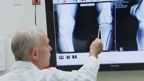
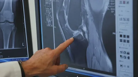
Explore the suite of advanced diagnostic tools available, such as smooth zooming, panning, and brightness/assessment changes.
Investigate precise tools for unique specialties, consisting as the Cobb Angle Tool for spinal checks in orthopedics.
Learn about the 3-D visualization competencies for viewing complex structures in extra detail.
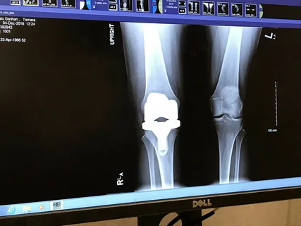
Understand how the viewer handles high-resolution snapshots from diverse modalities like CT, MRI, PET, and ultrasound.
-
Integration with AI:
In DICOM Onsite Model to Better Care, An investigation into how the AI-powered tools, along with anomaly detection and image evaluation, can be used within an on-site environment to assist with quicker and more accurate diagnoses.
Understand the AI’s function in areas like cardiology (echocardiograms) and oncology (early most cancers).
Next steps for targeted exploration
-
Request a demo:
Ask for an indication of the ALM DICOM Viewer, particularly on an on-site setup, to see the gear and capabilities in action within a neighborhood community environment.
-
Review case studies:
Look for case research or white papers from ALM Viewer that spotlight their onsite implementations in hospitals and clinics, focusing on the clinical and operational advantages.
-
Consult technical documentation:
Obtain technical specifications, machine requirements, and security documentation from ALM Viewer to apprehend the overall scope of the on-site version.
Key strategies for exploring the version
To discover the ALM DICOM onsite version for better care, attention on its key capabilities: cell and go-platform get entry to, AI-powered analysis, and secure, cloud-based storage. This aggregate improves collaboration, hastens analysis with gear like AI-assisted function detection and automatic analysis, and allows clinicians to access patient pix.
From any device, everywhere, to make more informed decisions quickly.
-
Leverage the cloud for seamless access:
Utilize the viewer’s cloud-primarily based architecture to get access to images from any place, whether in a medical institution, health center, or far-off exercise.
Access is to be had across more than one device, consisting of mobile systems, taking into consideration real-time image review and collaboration.
-
Integrate AI for quicker, greater accurate analysis:
Use the AI-powered radiology and pathology viewers to beautify photo processing and evaluation.
This equipment automates the detection of key functions and may highlight ability anomalies in scans like MRIs, CTs, and pathology pictures, lowering manual interpretation time and ability human errors.
-
Prioritize steady records management:
Confirm that the version uses a stable, cloud-based DICOM archive to guard sensitive patient information.
Ensure the machine is compliant with policies like HIPAA to hold patient privacy and information safety during transmission and storage.
-
Ensure pass-platform compatibility:
Verify that the PACS picture archiving viewer is compatible with all vital gadgets and operating systems used by your organization.
This flexibility guarantees that all authorized employees can get access to and analyze imaging information without being tied to unique hardware or locations.
-
Incorporate advanced photograph manipulation:
Utilize functions for comparison adjustment and zoom to enhance the visibility of diffused details in pics, making to more unique diagnostic paintings.
Explore 3-D rendering abilities for an extra comprehensive view of anatomical systems.
The Role of DICOM Viewer Onsite Model in Radiology
DICOM viewers are one of the maximum crucial equipment in current radiology, allowing for the green acquisition, manipulation, and interpretation of scientific images.
This equipment makes workflows less difficult, improves diagnostics, and integrates with imaging structures. Radiologists rely on DICOM viewers to view, analyze, and record findings with precision.
-
Image acquisition and retrieval:
DICOM viewers automatically fetch pics from imaging structures, which allows for very fast access to patient research. Advanced equipment guide pre-fetching and real-time streaming.
-
Enhanced Image Analysis and Editing:
In DICOM Onsite Model to Better Care, Features consisting of segmentation and dynamic assessment enhancement permit radiologists to mark anomalies and recognition attention to areas of interest.
-
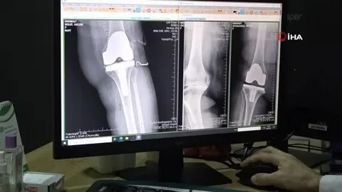
Support for Comparative Studies:
Radiologists can overlay or examine contemporary and past scans to monitor disease progression or treatment outcomes in DICOM Onsite Model to Better Care.
-
Error-Free Annotation and Reporting:
Annotation tools allow customers to mark findings, measure distances, and generate dependent reports, preserving consistency throughout studies.
-
Integration with PACS, RIS, and EHR:
Seamless integration allows coherent imaging statistics, updating patient facts, and an automated workflow.
Improved Collaboration and Communication: Immediately, photographs can be released for the collaboration of many specialists over telemedicine and remote analysis.
In short, the DICOM viewer streamlines radiology workflows, even as it removes mistakes and normally enhances patient care with its superior capabilities in managing and reading images.
The Beginner’s Guide to Using DICOM Viewer in OpenEMR
6 Features of DICOM Viewer
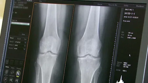
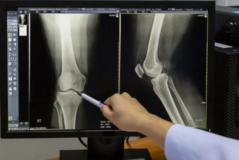
DICOM visitors offer particular tools like zoom, pan, brightness, evaluation changes, filters, and window leveling. These functionalities allow radiologists to decorate photographs, detect fractures, or pick out tumors by setting apart and enhancing critical visual information.
1. Tools for Image Enhancement and Adjustment
DICOM viewers offer specific gear like zoom, pan, brightness, evaluation changes, filters, and window leveling. These functionalities allow radiologists to decorate pix, detect fractures, or discover tumors by means of separating and improving crucial visual details.
2. 3-D Visualization and Volume Rendering Capabilities
Advanced DICOM viewers convert 2D slices into interactive 3D formats the using multiplanar reconstruction, volume rendering, and surface rendering. This equipment supplies special anatomical perspectives essential for oncology, neurosurgery, and surgical planning. So, companies can make certain correct measurements and higher scientific effects.
3. Seamless Integration with PACS, RIS, and DICOM
In DICOM Onsite Model to Better Care, Seamless PACS, RIS, and DICOM integration make certain bidirectional data flow, automatic document retrieval, and unified worklist management. Compliance with HIPAA requirements permits stable facts sharing and centralized access, streamlining workflows and enhancing diagnostic efficiency across radiology systems.
4. Annotation Features and Comprehensive Reporting
DICOM annotation gear guides specific measurements, text labels, arrows, and shapes. Features like macros, reporting templates, and image exports make certain standardized reporting and clear communication of findings for medical documentation, collaboration, and educational purposes.
5. Cross-platform compatibility and interoperability
DICOM Onsite Model to Better Care, ensure seamless compatibility across running structures and imaging modalities, inclusive of CT, MRI, PET, and ultrasound. API connectivity, adherence to DICOM standards, and cross-platform interoperability guide easy integration into various scientific workflows and research environments.
6. Advanced Tools for Image Comparison and Analysis
DICOM assessment tools allow synchronized scrolling, overlay options, and automatic alignment of multiple scans. This allows radiologists to investigate sickness development, examine remedy responses, and ensure accurate follow-ups, in particular in oncology, orthopedics, and longitudinal patient care.
DICOM Viewer Customization Service by way of CapMinds
Want to get the maximum out of the DICOM Viewer for your EMR systems?
We are a professional fitness tech organisation with years of experience in EHR, EMR, OpenEMR, HL7 FHIR, Mirth Connect, Health Interoperability, and More. Our DICOM customization carrier consists of:
Custom Layouts and Toolbars
Advanced Image Processing Capabilities
-
Comprehensive Data Visualization
In DICOM Onsite Model to Better Care, Our team of experts will work intently with you to tailor each component of the DICOM viewer in your EMR system, ensuring an easy workflow.
Elevate your diagnostic abilities of DICOM Onsite Model to Better Care and streamline your radiology practice with a DICOM viewer that honestly puts you in control.
Contact us these days and enjoy the full potential of the DICOM Onsite Model to Better Care in your Practice.
-
Faster Turnaround Times
In DICOM Onsite Model to Better Care, Time is often the difference between a high-quality and negative outcome in healthcare. DICOM permits quicker transmission and retrieval of scientific images, substantially decreasing prognosis turnaround times.
Traditional techniques, including bodily transporting film or CDs, can delay essential selections. However, DICOM gets rid of these delays.
A health facility in a smaller city can send an affected person’s CT test to a neuroradiologic in a metropolitan region within minutes. This speedy get right of entry to permits for fast evaluation, allowing faster interventions, which include thrombolysis or surgical treatment.
-
Improved Patient Outcomes
The mixture of greater accessibility and quicker turnaround instances without delay influences patient outcomes. DICOM presents timely diagnoses, which may be life-saving in many instances.
Quick get right of entry to to diagnostic imaging is vital, whether detecting a tumor early or diagnosing internal injuries after an accident.
For example, a patient in a rural health facility experiences sudden, excessive stomach pain. An ultrasound is dispatched to a radiologist through a DICOM Onsite Model to Better Care-enabled machine, who diagnoses appendicitis. This timely diagnosis allows the hospital to switch the patient to a surgical middle, stopping complications.
-
Cost-Effectiveness
Many small clinics and rural hospitals can’t always have the funds for to lease radiologists to work on-website online. DICOM-powered teleradiology gives a price-powerful answer: Doctors can study pix remotely. This offers facilities access to specialized understanding with no need for full-time personnel.
Besides, DICOM lowers operational prices by way of lowering the reliance on physical movie garage and shipping. Digital photos stored in PACS are less complicated to manage, retrieve, and proportion, saving money and time.
At our DICOM Onsite Model to Better Care, we’re devoted to creating answers that beautify care and make monetary sense for carriers. Our DICOM-enabled structures assist clinics and hospitals in stretching budgets without compromising patient care.
-
Scalable Solutions
The demand for clinical ALM medical imaging viewer demos is skyrocketing, driven by factors such as aging populations and advances in diagnostic imaging. DICOM guarantees that teleradiology structures can scale to meet this growing demand.
DICOM’s standardized framework helps the mixing of the latest imaging modalities, better record volumes, and increase healthcare companies’ networks.
A nearby hospital network can use a DICOM-primarily based teleradiology device to connect clinics, hospitals, and imaging facilities. The gadget can quickly grow as the community expands, permitting more customers, devices, and imaging studies.