ALM VIEWER

Key Components and Implementation of PACS

Key Components and Implementation of PACS in a US Prototype.

Introduction to PACS System

Picture Archiving and Communication System (PACS System) has become one of the most popular health care systems between 2003 and 2008. During this period, the media and interpretation media were replaced by the film based on digital imaging, which was considered an important success, where tools for digital images have become more well-known than the classic radiological traditions. Therefore, when a digital image of the chest is trapped, it can be treated directly by the computer.

Digital Imaging and PACS Integration

Digital imaging is a combination of PACS System machines and software hybrid systems used in digital imaging and communication to obtain, store, distribute, and reconstruct medical images using medicine (Dicom) standard. Through the integration of the Hospital Information System (HIS) and Radiology Information System (RIS), images and reports are digitally exchanged via PACs. This integration of PACS -RIS -HIS will end the need to store, restore, and view the movie jacket.

Importance in Healthcare

Previously, most of the healthcare system adopted the traditional way of storing and displaying patients’ data in hospitals, which delayed the time from imaging to reporting of interpretation. Providing medical goods information in a short time is an important step in relevant medical systems. As a result, the PACS System becomes crucial and ought to be used in hospitals to expedite physician assignments for patient care.

History and Development of PACS System

History and Development of PACS System

PACS System was first set out in the mid-1970s. Professor Jean-Raul introduced a medical information demonstration system, Dioogen, at Geneva University Hospitals, Switzerland. This system was later modernized to make PACs. At the moment, PAC has been adopted in many hospitals and medical institutions. In digital medical images, institutes could reduce the costs of data management and storage, and reduce time consumption in data transport. Pictures, as is known, are the basis for teaching medical imaging.

Therefore, it is important to provide students of medical imaging with high-quality images to improve the opportunity to analyze images. Based on this fact, a study was established to design the current PACS System with a medical imaging teaching method to create a better way to depict teaching systems in high institutions.

Challenges in Traditional Radiology Workflow

The most important boundaries of the traditional exam radiology cycle are as follows:

  • Time-consuming – determining clinical results cannot be achieved properly.
  • The physical recovery of films from the library can take hours, delaying treatment decisions.
  • The decision to refer to the doctor(s) varies from hours to days.
  • To save a copy of images, it is necessary to convert them to a digital format.
  • After installing the PACS System, the examination workflow is as follows:
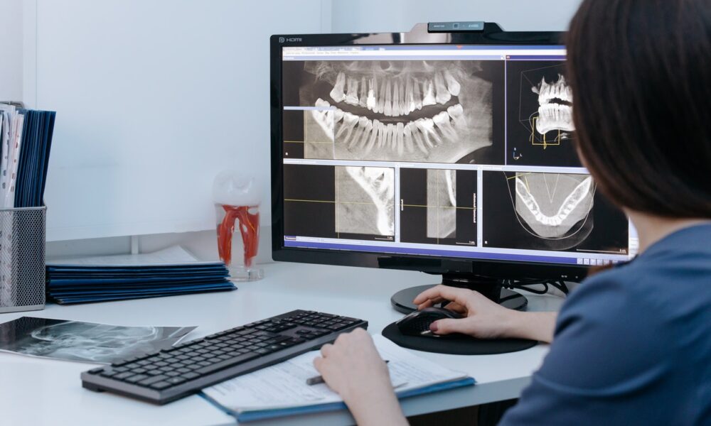
  • The technician takes digital images in the X-ray laboratory.
  • A few seconds later, the exposure was adjusted in the model workstation.
  • Images are then sent to digital archives.
  • Pictures are immediately available to radiologist (s), anywhere in the office and the Medical Institute of the Doctor (s).
PACS System Components

PACS component

Picture Archiving ​​and Communication System (PACS) consists of four main components: Image collection tool (Imaging Models), Communication Network and Servers, and Integrated Performance Workstation (WS). PACS communication networks allow PACs to be linked to RIS and their healthcare system.

Equipment for collecting images

Acquisition gateway computer equipment and depictions-terraces are tools used to acquire imagery.  Magnetic resonance imaging, computed tomography, PET, X-ray angiography, echocardiography, and other techniques are examples of image methods.  The Acquisition Gateway PC is used to interface with the Picture Archiving ​​and Communication System (PACS) server in this kind of fraud. The most important roles to use procurement port computers are to obtain images from imaging, convert the format to images from the manufacturer’s specification to the PACS standard format, called DICOM, and to perform some preaching features such as removing background and orientation to demonstrate any data pre -pre-processing functions.

Methods of Image Acquisition

There are two general ways to buy movies, photos, and digital procurement and digitalization. Digitization of regular films is an important way to convert radiological projections (movies) to digital images, as computers can only process digital images. Digitalizers such as laser scanners and charging units (CCD) can be used to achieve this image or film. The second method of image collection is the business of direct digital images, which can be done by using the time-developed X-Care equipment within the PACS System framework.

Benefits of Digital Image Collection

These devices can achieve digital images without the need for imaging plates used in traditional radiography. As a result, 30% of the image collection systems, including digital subtraction angiography, calculation tomography, magnetic resonance imaging, and ultrasound, are used to create digital images within the PACS System.

Communications network

Medical data can be sent between Picture Archiving ​​and Communication System (PACS) and other systems and components in distant places via the PACS communication network. Like other computer networks, PACS Network provides a route to communicate between the Imagine Modelity, Gateway Computer, PACS Server, Display and Review WSS, HIS/RIS system, and other external medical sites. 

Types of PACS Networks

Theoretically, main types of networks are used to transfer medical data on radiology:

  • LAN Network to connect various departments to a hospital (intra-hospital);
  • Tele-radiology network to transfer medical data to other external hospitals in that region.
  • Wide Area Network (WAN) – More extensive than a teleradiology network, a wide area network (WAN) links hospitals or medical institutions across cities, states, or even nations, allowing for remote image access and teleconsultation.

PACS Archive and Server

PACS Archive and Server

Information and imaging of all patients are sent to the PACS server for collection. The data collection is sent from the Gateway computer and from the PACS/RIS system to the PACS server. The Picture Archiving ​​and Communication System (PACS) server, which is the heart and engine of PACS, has two main components: storage media (database) and a collection system. The PAC collection system requires two levels for collection: short-term and long-term. Data (image) from a short-term level is restored in 2 seconds, while in the long term, people are restored in 3 minutes.

Storage Media Examples

  • RAID arrays for instant access to the most recent pictures
  • Magnetic drives to quickly retrieve reserved photos
  • Magneto-optic erasable disks for long-term storage
  • The library’s optical read-only memory
  • Modern flexible disks (DVD-RAM) for documents that can be expanded
  • Reinforcement tape with a high capacity

Important PACS Server Features

  • Use secure gateways to access photos.
  • Infrastructure is updated via database administration.
  • As pictures are transmitted, the display decides WSS.
  • Automatic image correlation from recorded tests
  • Conserve long-term library files or storage.
  • Automatically update advanced or registered radiography pictures
  • To display pictures, choose the brightness and complexity.

WSS – Workstation / Display

WSS – Workstation / Display

A presentation is a crucial component of the WS Picture Archiving ​​and Communication System (PACS) network that is necessary for clinical adoption of PAC. There is a hardware component that replaces the generator or manual light box in the radiology system. Today, most radiologists analyze films in a misleading room using light boxes or generators. Light boxes are bright boards, where ~ 12 films can be hung at once for review, and physical 8 of 200 films of a patient can be converted to a position to diagnose goals.

Simple image preparation operations such as zooming and film intervention are performed using a generator using an amplifier glass. The Display WSS helps the radiologist make a primary diagnosis, and they are therefore also designated as diagnostic WSS. WSS local storage database, communication network connections, resource management, and processing and performance software are its components. Basic imaging functions like access, manipulation, evaluation, and documentation are offered by Display WSS, making it a vital part of the PACS System. Secure on-premise image viewing is made possible by this integration with programs like the ALM DICOM Viewer On Site Model.

HL7 and DICOM standards

Two factors make it challenging to broadcast pictures and reports between various medical facilities: First, medical information and paintings are created using various image kinds from particular manufacturers, and second, the information system uses various machine platforms. In order to address this, the ALM DICOM Viewer Cloud Model and ALM DICOM Viewer Hybrid Model guarantee smooth compatibility with DICOM and HL7 standards.

Health condition 7 (HL7) and DICOM, asymmetrical images of various restorations are being processed in a composition system, along with rising medical requirements. Two elements are required to interface two medical systems: a general data format and a correspondence protocol, both of which are supported within a PACS System to ensure seamless interoperability.

HL7 Standard

HL7 is a standard text-based information format, and DICOM covers data format and correspondence protocol. In accordance with the 10 HL7 standards, DICOM is understandable to exchange medical information such as HIS, RIS, and a PACS System. By adjusting the DICOM standard, medical images created by classification of types and manufacturers can be integrated into a healthcare system, making the PACS System a vital component for seamless data sharing and management.

HL7 was introduced in March 1987 to create a standard for electronic information trading between a customer and supplier, especially to create a standard for hospital regulations and electronic information trading. At the highest level, HL7 Standard operates at the determination level, at the range level in the seven communication levels of Open System Interconnection (OSI). The main goal is to improve the execution of the interface between PC regulations from different manufacturers, ensuring smooth integration with systems like the PACS System for better medical data management.

DICOM Standard

It confirms the exchange of data between the standard health care systems, such as HIS, RIS, and PACS systems. On the other hand, DICOM is an important standard, which was developed in 1993 as a result of early diligence by the ACR and NEMA Joint Council to continue correspondence with computer-controlled image data regardless of the imaging manufacturer. This standard encourages progress and development of the PACS System for interfacing with different treatment center data systems in a uniform way. In addition, DICOM allows a symbolic database that can be cross-influenced by a broad classification of geographically expressed things.

In 2008, a task discussed the integration of a research called content-based image collection with RIS and PACS systems, which aims to improve the workflow in the radiological daily routine. The importance of this integration comes from offering all PAC collections for radiologists to make an accurate diagnosis for the current study of patients. In this article, integration between RIS and PACS System is achieved in Webxas, where the study (image and information) for all patients is stored in the server database, and Webxas can call any study using the patient’s name and the number of studies. Experts can then portray images of a specific study and analyze them for better diagnosis from the current study.

The name of the ACR, officially known as the American College of Radiology, and the national electric manufacturer, made a selection to create a set of benchmarks to act as a general background for various medical imaging providers. The main goal was that the recent equipment would have the opportunity to participate in and especially within the PACS System domain. The Committee, which was primarily focused on issues related to data exchange, connections, and communication between the healthcare system, began work in 1982.

Webxa-Prototype application

Webxa-Prototype application

Webxa Picture Archiving ​​and Communication System (PACS) is the prototype application that has a similar structural design to the client-server, except that the client and server have software-based applications. The extra benefits of the online design server on the client server are as follows: First, customers’ WS equipment can be independent as long as the web software is outlined. Second, web-based software runs perfectly; that is, web-based applications can be used in any location until an Internet connection is provided.

The disadvantage of online applications is that their functionality and performance are limited compared to client-server applications. One of the most important purposes of using online public applications in the health care system is real-time telecommunications or telecommunications. Advanced Zero Trust Security is another advantage of the web-based architecture, which protects medical imaging data during online cooperation and teleconsultations..

Teleconsultation

Teleconsultation deals with various scenarios in health institutions. Let’s consider the following scenario: When a doctor in the medical department wants to consult a radiologist in a reader’s reader, about a patient’s written report in the check room. This counseling process, both experts, should be without leaving their departments and running in every way to each other’s departments. Therefore, the PACS System enables consultation, a situation where two or more experts in different departments need to discuss and consult the patient’s results without leaving their departments.

Previously, telecommunications tools such as Televidio and networking tools had some consequences, which are a Microsoft PCANYWHERE sharing software from Intel, and PCANYWHERE from Intel and Cementc. However, most of these applications are deficient in explaining the purpose of image processing functions required in the healthcare system, which highlights the importance of adopting a PACS System for efficient image handling and communication.

In this letter, the Online Audience app, called Webxa, is made on PAC and tested. This application is only installed and established for primary clinical purposes, where medical imaging software is integrated with communication functions to develop an external view and consultation equipment using TCP/IP protocols, further enhancing the efficiency of a PACS System in clinical practice.

Real-Time Insights and Interactive PACS Features

A real-time text exchange property is added to the WebxA application to make it easier for experts to exchange comments and consult patients’ reports and results.

Simple and friendly GUI has buttons to stop the frame and move forward or backward due to angiography. The text exchange scene can be used by experts to exchange comments on the primary diagnosis of medical images or patients’ data, making the PACS System more interactive and collaborative.

Client/Picture Archiving ​​and Communication System (PACS)  Server communication through TCP/IP.

Software features such as storage, open, and print.